protein K33-linked deubiquitination / negative regulation of proteasomal ubiquitin-dependent protein catabolic process / negative regulation of innate immune response / protein sequestering activity / regulation of protein stability / ubiquitin-dependent protein catabolic process / cysteine-type deubiquitinase activity / ubiquitinyl hydrolase 1 / nucleus / cytoplasm / cytosol Similarity search - Function
Mass: 18.015 Da / Num. of mol.: 221 / Source method: isolated from a natural source / Formula: H2O
Has protein modification
Y
-
Experimental details
-
Experiment
Experiment
Method: X-RAY DIFFRACTION / Number of used crystals: 1
-
Sample preparation
Crystal
Density Matthews: 2.92 Å3/Da / Density % sol: 57.92 %
Crystal grow
Temperature: 291 K / Method: vapor diffusion, hanging drop / pH: 7.5 Details: Protein sample was first mixed Hampton Research Additive Screen H03 40% in 10:1 (V/V) Protein:1,3-propanediol ratio, then set up with 15% PEG8000, 0.2 M MgCl2, 0.1 M HEPES, pH 7.5, vapor ...Details: Protein sample was first mixed Hampton Research Additive Screen H03 40% in 10:1 (V/V) Protein:1,3-propanediol ratio, then set up with 15% PEG8000, 0.2 M MgCl2, 0.1 M HEPES, pH 7.5, vapor diffusion, hanging drop, temperature 291K, VAPOR DIFFUSION, HANGING DROP
Protocol: SINGLE WAVELENGTH / Monochromatic (M) / Laue (L): M / Scattering type: x-ray
Radiation wavelength
Wavelength: 0.97932 Å / Relative weight: 1
Reflection
Resolution: 2.06→50 Å / Num. obs: 37072 / % possible obs: 99.8 % / Redundancy: 9.7 % / Biso Wilson estimate: 29.4 Å2 / Rmerge(I) obs: 0.127 / Χ2: 1.463 / Net I/σ(I): 22.9
Reflection shell
Resolution (Å)
Redundancy (%)
Rmerge(I) obs
Num. unique all
Χ2
Diffraction-ID
% possible all
2.06-2.1
5.9
0.82
1797
0.625
1
97.3
2.1-2.13
6.8
0.737
1791
0.674
1
99.2
2.13-2.17
8
0.735
1807
0.645
1
100
2.17-2.22
9.1
0.679
1868
0.67
1
100
2.22-2.27
9.5
0.593
1791
0.707
1
100
2.27-2.32
9.9
0.535
1842
0.719
1
100
2.32-2.38
10.2
0.446
1822
0.753
1
100
2.38-2.44
10.5
0.41
1808
0.77
1
100
2.44-2.51
10.5
0.354
1855
0.806
1
100
2.51-2.6
10.6
0.301
1828
0.855
1
100
2.6-2.69
10.5
0.269
1847
0.905
1
100
2.69-2.8
10.6
0.226
1841
1
1
100
2.8-2.92
10.4
0.179
1839
1.101
1
100
2.92-3.08
10.4
0.144
1866
1.242
1
100
3.08-3.27
10.4
0.121
1855
1.388
1
100
3.27-3.52
10.3
0.095
1872
1.756
1
100
3.52-3.88
10.2
0.084
1877
2.281
1
100
3.88-4.44
10
0.08
1887
3.09
1
100
4.44-5.59
9.8
0.078
1919
3.269
1
100
5.59-50
9.4
0.076
2060
4.91
1
100
-
Processing
Software
Name
Version
Classification
NB
SCALEPACK
datascaling
REFMAC
5.8.0073
refinement
PDB_EXTRACT
3.15
dataextraction
SBC-Collect
datacollection
HKL-3000
datareduction
HKL-3000
datascaling
SOLVE
phasing
RESOLVE
phasing
Refinement
Method to determine structure: SAD / Resolution: 2.06→50 Å / Cor.coef. Fo:Fc: 0.96 / Cor.coef. Fo:Fc free: 0.927 / WRfactor Rfree: 0.2152 / WRfactor Rwork: 0.1719 / FOM work R set: 0.8246 / SU B: 4.505 / SU ML: 0.119 / SU R Cruickshank DPI: 0.1641 / SU Rfree: 0.1612 / Cross valid method: THROUGHOUT / σ(F): 0 / ESU R: 0.164 / ESU R Free: 0.161 / Stereochemistry target values: MAXIMUM LIKELIHOOD / Details: HYDROGENS HAVE BEEN ADDED IN THE RIDING POSITIONS
Rfactor
Num. reflection
% reflection
Selection details
Rfree
0.2419
1137
3.1 %
RANDOM
Rwork
0.1891
-
-
-
obs
0.1906
35715
99.84 %
-
Solvent computation
Ion probe radii: 0.8 Å / Shrinkage radii: 0.8 Å / VDW probe radii: 1.2 Å / Solvent model: MASK
In the structure databanks used in Yorodumi, some data are registered as the other names, "COVID-19 virus" and "2019-nCoV". Here are the details of the virus and the list of structure data.
Jan 31, 2019. EMDB accession codes are about to change! (news from PDBe EMDB page)
EMDB accession codes are about to change! (news from PDBe EMDB page)
The allocation of 4 digits for EMDB accession codes will soon come to an end. Whilst these codes will remain in use, new EMDB accession codes will include an additional digit and will expand incrementally as the available range of codes is exhausted. The current 4-digit format prefixed with “EMD-” (i.e. EMD-XXXX) will advance to a 5-digit format (i.e. EMD-XXXXX), and so on. It is currently estimated that the 4-digit codes will be depleted around Spring 2019, at which point the 5-digit format will come into force.
The EM Navigator/Yorodumi systems omit the EMD- prefix.
Related info.:Q: What is EMD? / ID/Accession-code notation in Yorodumi/EM Navigator
Yorodumi is a browser for structure data from EMDB, PDB, SASBDB, etc.
This page is also the successor to EM Navigator detail page, and also detail information page/front-end page for Omokage search.
The word "yorodu" (or yorozu) is an old Japanese word meaning "ten thousand". "mi" (miru) is to see.
Related info.:EMDB / PDB / SASBDB / Comparison of 3 databanks / Yorodumi Search / Aug 31, 2016. New EM Navigator & Yorodumi / Yorodumi Papers / Jmol/JSmol / Function and homology information / Changes in new EM Navigator and Yorodumi
 Movie
Movie Controller

 Yorodumi
 Open data
 Basic information
 Components
 Keywords
 Function and homology information
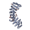
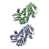
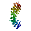
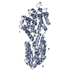
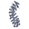
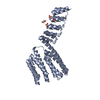
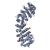
 Homo sapiens (human)
 X-RAY DIFFRACTION /
  Authors
 Citation
 Structure visualization
 Downloads & links
 Other downloads
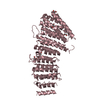


 PDBj
PDBj Assembly


 Mass: 35.453 Da / Num. of mol.: 1 / Source method: obtained synthetically / Formula: Cl
 Mass: 62.068 Da / Num. of mol.: 1 / Source method: obtained synthetically / Formula: C2H6O2
 Num. of mol.: 5 / Source method: obtained synthetically
 Mass: 18.015 Da / Num. of mol.: 221 / Source method: isolated from a natural source / Formula: H2O
 Sample preparation
 / Beamline: 19-ID / Wavelength: 0.97932 Å
 Processing