 Movie
Movie Controller

[English] 日本語
 Yorodumi
- PDB-4rpt: The 1.35A structure of a viral RNase L antagonist reveals basis f... -
+ Open data

- Basic information
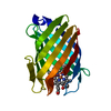
| Entry | Database: PDB / ID: 4rpt | ||||||
|---|---|---|---|---|---|---|---|
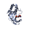
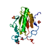
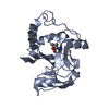
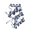
| Title | The 1.35A structure of a viral RNase L antagonist reveals basis for the 2'-5'-oligoadenylate binding and enzyme activity. | ||||||
 Components Components | Capping enzyme protein | ||||||
 Keywords Keywords | VIRAL PROTEIN / 2H phosphodiesterase | ||||||
| Function / homology |  Function and homology information Function and homology informationHydrolases; Acting on ester bonds; Phosphoric-diester hydrolases / phosphoric diester hydrolase activity / viral process / viral nucleocapsid / mRNA guanylyltransferase / mRNA guanylyltransferase activity / mRNA (guanine-N7)-methyltransferase / mRNA 5'-cap (guanine-N7-)-methyltransferase activity / symbiont-mediated suppression of host innate immune response / GTP binding / RNA binding Similarity search - Function | ||||||
| Biological species |  Rotavirus A Rotavirus A | ||||||
| Method |  X-RAY DIFFRACTION / SYNCHROTRON / SAD / Resolution: 1.35 Å X-RAY DIFFRACTION / SYNCHROTRON / SAD / Resolution: 1.35 Å | ||||||
 Authors Authors | Hu, L. / Prasad, B.V.V. | ||||||
 Citation Citation | Journal: J.Virol. / Year: 2015 Title: Structural basis for 2'-5'-oligoadenylate binding and enzyme activity of a viral RNase L antagonist. Authors: Ogden, K.M. / Hu, L. / Jha, B.K. / Sankaran, B. / Weiss, S.R. / Silverman, R.H. / Patton, J.T. / Prasad, B.V. | ||||||
| History |
|
- Structure visualization
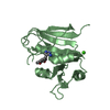
| Structure viewer | Molecule: MolmilJmol/JSmol |
|---|
- Downloads & links
-Download
| PDBx/mmCIF format | 4rpt.cif.gz | 77.6 KB | Display | PDBx/mmCIF format |
|---|---|---|---|---|
| PDB format | pdb4rpt.ent.gz | 58.4 KB | Display | PDB format |
| PDBx/mmJSON format | 4rpt.json.gz | Tree view | PDBx/mmJSON format | |
| Others |  Other downloads Other downloads |
-Validation report
| Arichive directory | https://data.pdbj.org/pub/pdb/validation_reports/rp/4rptftp://data.pdbj.org/pub/pdb/validation_reports/rp/4rpt | HTTPS FTP |
|---|
-Related structure data


-Links
 PDBj
PDBj


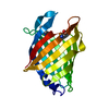
- Assembly
| Deposited unit | 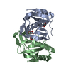
| ||||||||
|---|---|---|---|---|---|---|---|---|---|
| 1 | 
| ||||||||
| 2 | 
| ||||||||
| 3 |
| ||||||||
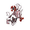
| Unit cell |
|
-Components
| #1: Protein | Mass: 16766.479 Da / Num. of mol.: 2 / Fragment: C-terminal domain, UNP residues 694-835 Source method: isolated from a genetically manipulated source Source: (gene. exp.) Rotavirus A / Strain: RRV / Production host:  #2: Chemical |   Mass: 102.046 Da / Num. of mol.: 2 / Source method: obtained synthetically / Formula: C3H2O4 Mass: 102.046 Da / Num. of mol.: 2 / Source method: obtained synthetically / Formula: C3H2O4#3: Chemical |   Mass: 62.068 Da / Num. of mol.: 2 / Source method: obtained synthetically / Formula: C2H6O2 Mass: 62.068 Da / Num. of mol.: 2 / Source method: obtained synthetically / Formula: C2H6O2#4: Water | ChemComp-HOH / |  Mass: 18.015 Da / Num. of mol.: 328 / Source method: isolated from a natural source / Formula: H2O Mass: 18.015 Da / Num. of mol.: 328 / Source method: isolated from a natural source / Formula: H2O |
|---|
-Experimental details
-Experiment
| Experiment | Method: X-RAY DIFFRACTION / Number of used crystals: 1 |
|---|
- Sample preparation
| Crystal | Density Matthews: 2.06 Å3/Da / Density % sol: 40.23 % |
|---|---|
| Crystal grow | Temperature: 298 K / pH: 4 Details: 14% PEG3350, 0.2 M sodium malonate, pH 4, 1 M NaI, VAPOR DIFFUSION, HANGING DROP, temperature 298.0K |
-Data collection
| Diffraction | Mean temperature: 95 K |
|---|---|
| Diffraction source | Source: SYNCHROTRON / Site: ALS  / Beamline: 5.0.1 / Wavelength: 0.9774 / Beamline: 5.0.1 / Wavelength: 0.9774 |
| Detector | Type: ADSC QUANTUM 315r / Detector: CCD / Date: Apr 28, 2014 |
| Radiation | Monochromator: SI(220) ASYMMETRIC CUT SINGLE CRYSTAL / Protocol: SINGLE WAVELENGTH / Monochromatic (M) / Laue (L): M / Scattering type: x-ray |
| Radiation wavelength | Wavelength: 0.9774 Å / Relative weight: 1 |
| Reflection | Resolution: 1.35→31.86 Å / Num. obs: 56612 / % possible obs: 95.2 % / Observed criterion σ(I): 2 / Redundancy: 4.9 % / Rmerge(I) obs: 0.039 / Rsym value: 0.044 / Net I/σ(I): 19.2 |
| Reflection shell | Resolution: 1.35→1.42 Å / Rmerge(I) obs: 0.471 / Mean I/σ(I) obs: 3.2 / Rsym value: 0.528 / % possible all: 93.5 |
- Processing
| Software |
| |||||||||||||||||||||||||||||||||||||||||||||||||||||||||||||||||||||||||||||||||||||||||||||||||||||||||||||||||||||||||||||||||||||||||||||||||||||||||||||||||||||||||||||||||||||||||||||||||||||||||||||||||||||||||
|---|---|---|---|---|---|---|---|---|---|---|---|---|---|---|---|---|---|---|---|---|---|---|---|---|---|---|---|---|---|---|---|---|---|---|---|---|---|---|---|---|---|---|---|---|---|---|---|---|---|---|---|---|---|---|---|---|---|---|---|---|---|---|---|---|---|---|---|---|---|---|---|---|---|---|---|---|---|---|---|---|---|---|---|---|---|---|---|---|---|---|---|---|---|---|---|---|---|---|---|---|---|---|---|---|---|---|---|---|---|---|---|---|---|---|---|---|---|---|---|---|---|---|---|---|---|---|---|---|---|---|---|---|---|---|---|---|---|---|---|---|---|---|---|---|---|---|---|---|---|---|---|---|---|---|---|---|---|---|---|---|---|---|---|---|---|---|---|---|---|---|---|---|---|---|---|---|---|---|---|---|---|---|---|---|---|---|---|---|---|---|---|---|---|---|---|---|---|---|---|---|---|---|---|---|---|---|---|---|---|---|---|---|---|---|---|---|---|---|
| Refinement | Method to determine structure: SAD / Resolution: 1.35→31.86 Å / SU ML: 0.16 / σ(F): 2 / Phase error: 23.41 / Stereochemistry target values: ML
| |||||||||||||||||||||||||||||||||||||||||||||||||||||||||||||||||||||||||||||||||||||||||||||||||||||||||||||||||||||||||||||||||||||||||||||||||||||||||||||||||||||||||||||||||||||||||||||||||||||||||||||||||||||||||
| Solvent computation | Shrinkage radii: 0.9 Å / VDW probe radii: 1.11 Å / Solvent model: FLAT BULK SOLVENT MODEL | |||||||||||||||||||||||||||||||||||||||||||||||||||||||||||||||||||||||||||||||||||||||||||||||||||||||||||||||||||||||||||||||||||||||||||||||||||||||||||||||||||||||||||||||||||||||||||||||||||||||||||||||||||||||||
| Refinement step | Cycle: LAST / Resolution: 1.35→31.86 Å
| |||||||||||||||||||||||||||||||||||||||||||||||||||||||||||||||||||||||||||||||||||||||||||||||||||||||||||||||||||||||||||||||||||||||||||||||||||||||||||||||||||||||||||||||||||||||||||||||||||||||||||||||||||||||||
| Refine LS restraints |
| |||||||||||||||||||||||||||||||||||||||||||||||||||||||||||||||||||||||||||||||||||||||||||||||||||||||||||||||||||||||||||||||||||||||||||||||||||||||||||||||||||||||||||||||||||||||||||||||||||||||||||||||||||||||||
| LS refinement shell |
|
