D res high: 2 Å / D res low: 50 Å / FOM : 0 / FOM acentric: 0.497 / FOM centric: 0 / Reflection: 0 / Reflection acentric: 18792 / Reflection centric: 0
Phasing MAD set
R cullis centric: 0 / Highest resolution: 2 Å / Lowest resolution: 50 Å / Loc centric: 0 / Power centric: 0 / Reflection centric: _
ID
R cullis acentric
Loc acentric
Power acentric
Reflection acentric
1
1.32
0.1
0
18792
2
0.94
3.7
0.75
18707
Phasing MAD set shell
R cullis centric: 0 / Loc centric: 0 / Power centric: 0 / Reflection centric: _
ID
Resolution (Å)
R cullis acentric
Loc acentric
Power acentric
Reflection acentric
1
12.5-50
1.11
0.2
0
42
1
7.14-12.5
1.24
0.2
0
298
1
5-7.14
1.28
0.2
0
794
1
3.85-5
1.04
0.2
0
1441
1
3.13-3.85
1.03
0.1
0
2276
1
2.63-3.13
1.43
0.1
0
3361
1
2.27-2.63
1.7
0.1
0
4555
1
2-2.27
1.91
0
0
6025
2
12.5-50
1.05
19.3
0.42
41
2
7.14-12.5
1.01
10.4
0.84
284
2
5-7.14
0.91
7.3
1.01
779
2
3.85-5
0.94
6.8
0.83
1408
2
3.13-3.85
0.92
5.3
0.8
2260
2
2.63-3.13
0.91
3.5
0.83
3355
2
2.27-2.63
0.93
2.7
0.71
4555
2
2-2.27
0.98
2.3
0.52
6025
Phasing MAD set site
Atom type symbol: Se
ID
B iso
Fract x
Fract y
Fract z
Occupancy
Occupancy iso
1
32.0739
-0.004
-0.044
-0.125
5.638
0
2
35.9926
-0.395
0.278
-0.254
5.085
0
3
37.6005
-0.004
-0.044
-0.126
4.241
-0.148
4
37.8912
-0.395
0.278
-0.254
3.558
-0.126
Phasing MAD shell
Resolution (Å)
FOM
FOM acentric
FOM centric
Reflection
Reflection acentric
Reflection centric
12.5-50
0
0.212
0
1484
42
0
7.14-12.5
0
0.37
0
298
0
5-7.14
0
0.624
0
5349136
794
0
3.85-5
0
0.52
0
1441
0
3.13-3.85
0
0.553
0
2276
0
2.63-3.13
0
0.587
0
3361
0
2.27-2.63
0
0.533
0
1053855744
4555
0
2-2.27
0
0.386
0
10980
6025
0
Phasing dm
Method: Solvent flattening and Histogram matching / Reflection: 28121
Phasing dm shell
Resolution (Å)
Delta phi final
FOM
Reflection
6.43-100
53.8
0.832
501
5.18-6.43
41.3
0.93
508
4.55-5.18
46.1
0.943
522
4.1-4.55
46.4
0.947
588
3.76-4.1
50.2
0.949
630
3.5-3.76
51.6
0.952
692
3.28-3.5
45.6
0.948
736
3.1-3.28
49.3
0.941
798
2.95-3.1
45
0.943
824
2.82-2.95
45.7
0.942
870
2.7-2.82
45.1
0.938
940
2.6-2.7
44.1
0.936
915
2.5-2.6
46.5
0.942
1015
2.42-2.5
47
0.94
1018
2.35-2.42
47.6
0.937
1045
2.28-2.35
50.4
0.934
1085
2.21-2.28
52
0.933
1137
2.16-2.21
51.5
0.927
1148
2.1-2.16
54.8
0.924
1217
2.05-2.1
58.7
0.925
1207
2.01-2.05
60
0.914
1238
1.96-2.01
85.7
0.919
1270
1.92-1.96
88.7
0.91
1286
1.88-1.92
90.9
0.881
1302
1.85-1.88
91
0.872
1376
1.81-1.85
88.6
0.859
1389
1.78-1.81
92.3
0.781
1363
1.75-1.78
90
0.612
1501
-
Processing
Software
Name
Version
Classification
NB
DENZO
datareduction
SCALEPACK
datascaling
MLPHARE
phasing
DM
6.1
phasing
REFMAC
5.8.0073
refinement
PDB_EXTRACT
3.15
dataextraction
SBC-Collect
datacollection
HKL-3000
datareduction
HKL-3000
SHELXD
phasing
SHELXE
modelbuilding
SOLVE
phasing
RESOLVE
phasing
ARP/wARP
modelbuilding
CCP4
phasing
O
modelbuilding
Coot
modelbuilding
Refinement
Method to determine structure: MAD / Resolution: 1.75→29.31 Å / Cor.coef. Fo:Fc: 0.968 / Cor.coef. Fo:Fc free: 0.957 / WRfactor Rfree: 0.2094 / WRfactor Rwork: 0.1752 / FOM work R set: 0.9094 / SU B: 4.086 / SU ML: 0.067 / SU R Cruickshank DPI: 0.1062 / SU Rfree: 0.1025 / Cross valid method: THROUGHOUT / σ(F): 0 / ESU R: 0.106 / ESU R Free: 0.102 Stereochemistry target values: MAXIMUM LIKELIHOOD WITH PHASES Details: HYDROGENS HAVE BEEN ADDED IN THE RIDING POSITIONS
Rfactor
Num. reflection
% reflection
Selection details
Rfree
0.1976
1421
5.1 %
RANDOM
Rwork
0.1665
-
-
-
obs
0.1681
26698
99.65 %
-
all
-
26698
-
-
Solvent computation
Ion probe radii: 0.8 Å / Shrinkage radii: 0.8 Å / VDW probe radii: 1.2 Å / Solvent model: MASK
In the structure databanks used in Yorodumi, some data are registered as the other names, "COVID-19 virus" and "2019-nCoV". Here are the details of the virus and the list of structure data.
Jan 31, 2019. EMDB accession codes are about to change! (news from PDBe EMDB page)
EMDB accession codes are about to change! (news from PDBe EMDB page)
The allocation of 4 digits for EMDB accession codes will soon come to an end. Whilst these codes will remain in use, new EMDB accession codes will include an additional digit and will expand incrementally as the available range of codes is exhausted. The current 4-digit format prefixed with “EMD-” (i.e. EMD-XXXX) will advance to a 5-digit format (i.e. EMD-XXXXX), and so on. It is currently estimated that the 4-digit codes will be depleted around Spring 2019, at which point the 5-digit format will come into force.
The EM Navigator/Yorodumi systems omit the EMD- prefix.
Related info.:Q: What is EMD? / ID/Accession-code notation in Yorodumi/EM Navigator
Yorodumi is a browser for structure data from EMDB, PDB, SASBDB, etc.
This page is also the successor to EM Navigator detail page, and also detail information page/front-end page for Omokage search.
The word "yorodu" (or yorozu) is an old Japanese word meaning "ten thousand". "mi" (miru) is to see.
Related info.:EMDB / PDB / SASBDB / Comparison of 3 databanks / Yorodumi Search / Aug 31, 2016. New EM Navigator & Yorodumi / Yorodumi Papers / Jmol/JSmol / Function and homology information / Changes in new EM Navigator and Yorodumi
 Movie
Movie Controller

 Yorodumi
 Open data
 Basic information
 Components
 Keywords
 Function and homology information
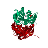
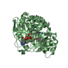
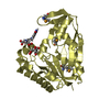
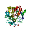
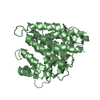
 Salmonella enterica subsp. enterica serovar Typhimurium str. 14028S (bacteria)
 X-RAY DIFFRACTION /
  Authors
 Citation
 Structure visualization
 Downloads & links
 Other downloads


 PDBj
PDBj


 Assembly
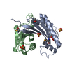


 Mass: 96.063 Da / Num. of mol.: 6 / Source method: obtained synthetically / Formula: SO4
 Mass: 118.174 Da / Num. of mol.: 1 / Source method: obtained synthetically / Formula: C6H14O2 / Comment: precipitant*YM
 Mass: 60.052 Da / Num. of mol.: 1 / Source method: obtained synthetically / Formula: C2H4O2
 Mass: 18.015 Da / Num. of mol.: 192 / Source method: isolated from a natural source / Formula: H2O
 Sample preparation
 / Beamline: 19-ID / Wavelength: 0.97915, 0.97929
 Processing