 Movie
Movie Controller

[English] 日本語
 Yorodumi
- PDB-4r80: Crystal Structure of a De Novo Designed Beta Sheet Protein, Cysta... -
+ Open data

- Basic information
| Entry | Database: PDB / ID: 4r80 | ||||||
|---|---|---|---|---|---|---|---|
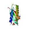
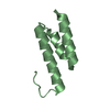
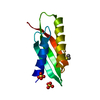
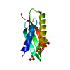
| Title | Crystal Structure of a De Novo Designed Beta Sheet Protein, Cystatin Fold, Northeast Structural Genomics Consortium (NESG) Target OR486 | ||||||
 Components Components | OR486 | ||||||
 Keywords Keywords | DE NOVO PROTEIN / Structural Genomics / PSI-Biology / Protein Structure Initiative / Northeast Structural Genomics Consortium (NESG) / Target OR486 / protein engineering / denovo beta sheet design / Cystatin Fold | ||||||
| Function / homology | Nuclear Transport Factor 2; Chain: A, - #630 / Nuclear Transport Factor 2; Chain: A, / Roll / Alpha Beta Function and homology information Function and homology information | ||||||
| Biological species | synthetic construct (others) | ||||||
| Method |  X-RAY DIFFRACTION / SYNCHROTRON / MOLECULAR REPLACEMENT / Resolution: 2.445 Å X-RAY DIFFRACTION / SYNCHROTRON / MOLECULAR REPLACEMENT / Resolution: 2.445 Å | ||||||
 Authors Authors | Guan, R. / Marcos, E. / O'Connell, P. / Seetharaman, J. / Janjua, H. / Xiao, R. / Maglaqui, M. / Acton, T.B. / Everett, J.K. / Baker, D. ...Guan, R. / Marcos, E. / O'Connell, P. / Seetharaman, J. / Janjua, H. / Xiao, R. / Maglaqui, M. / Acton, T.B. / Everett, J.K. / Baker, D. / Montelione, G.T. / Northeast Structural Genomics Consortium (NESG) | ||||||
 Citation Citation | Journal: To be Published Title: Crystal Structure of an engineered protein with denovo beta sheet design, Northeast Structural Genomics Consortium (NESG) Target OR486 Authors: Guan, R. / Marcos, E. / O'Connell, P. / Seetharaman, J. / Janjua, H. / Xiao, R. / Maglaqui, M. / Acton, T.B. / Everett, J.K. / Baker, D. / Montelione, G.T. / Northeast Structural Genomics Consortium (NESG) | ||||||
| History |
|
- Structure visualization
| Structure viewer | Molecule: MolmilJmol/JSmol |
|---|
- Downloads & links
-Download
| PDBx/mmCIF format | 4r80.cif.gz | 41.8 KB | Display | PDBx/mmCIF format |
|---|---|---|---|---|
| PDB format | pdb4r80.ent.gz | 30.6 KB | Display | PDB format |
| PDBx/mmJSON format | 4r80.json.gz | Tree view | PDBx/mmJSON format | |
| Others |  Other downloads Other downloads |
-Validation report
| Arichive directory | https://data.pdbj.org/pub/pdb/validation_reports/r8/4r80ftp://data.pdbj.org/pub/pdb/validation_reports/r8/4r80 | HTTPS FTP |
|---|
-Related structure data
| Similar structure data | |
|---|---|
| Other databases |


-Links
 PDBj
PDBj
- Assembly
| Deposited unit | 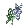
| ||||||||||||
|---|---|---|---|---|---|---|---|---|---|---|---|---|---|
| 1 | 
| ||||||||||||
| 2 | 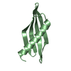
| ||||||||||||
| Unit cell |
| ||||||||||||
| Components on special symmetry positions |
| ||||||||||||
| Details | The biological molecule is unknown |
-Components
| #1: Protein | Mass: 9897.169 Da / Num. of mol.: 2 Source method: isolated from a genetically manipulated source Source: (gene. exp.) synthetic construct (others) / Production host:  #2: Water | ChemComp-HOH / |  Mass: 18.015 Da / Num. of mol.: 59 / Source method: isolated from a natural source / Formula: H2O Mass: 18.015 Da / Num. of mol.: 59 / Source method: isolated from a natural source / Formula: H2O |
|---|
-Experimental details
-Experiment
| Experiment | Method: X-RAY DIFFRACTION / Number of used crystals: 1 |
|---|
- Sample preparation
| Crystal | Density Matthews: 1.89 Å3/Da / Density % sol: 34.9 % |
|---|---|
| Crystal grow | Temperature: 295 K / Method: vapor diffusion, hanging drop / pH: 5.5 Details: Protein solution: 100mM NaCl, 5mM DTT, 0.02% NaN3, 10mM Tris-HCl (pH 7.5), 10mg/ml. Reservoir solution:0.1 M NaH2PO4, 0.1 M Na Acetate, pH 5.5, 28% PEG 400, VAPOR DIFFUSION, HANGING DROP, temperature 295K |
-Data collection
| Diffraction | Mean temperature: 100 K |
|---|---|
| Diffraction source | Source: SYNCHROTRON / Site: NSLS  / Beamline: X4C / Wavelength: 0.97916 Å / Beamline: X4C / Wavelength: 0.97916 Å |
| Detector | Type: MAR CCD 165 mm / Detector: CCD / Date: Aug 11, 2014 |
| Radiation | Monochromator: Si 111 CHANNEL / Protocol: SINGLE WAVELENGTH / Monochromatic (M) / Laue (L): M / Scattering type: x-ray |
| Radiation wavelength | Wavelength: 0.97916 Å / Relative weight: 1 |
| Reflection | Resolution: 2.445→41.671 Å / Num. all: 5311 / Num. obs: 5311 / % possible obs: 93.1 % / Observed criterion σ(F): 2 / Observed criterion σ(I): 2 / Redundancy: 2.3 % / Rmerge(I) obs: 0.052 / Net I/σ(I): 33.87 |
| Reflection shell | Resolution: 2.44→2.48 Å / Redundancy: 1.4 % / Rmerge(I) obs: 0.06 / % possible all: 42.8 |
- Processing
| Software |
| ||||||||||||||||||||||||||||||||||||||||||||||||||||||||
|---|---|---|---|---|---|---|---|---|---|---|---|---|---|---|---|---|---|---|---|---|---|---|---|---|---|---|---|---|---|---|---|---|---|---|---|---|---|---|---|---|---|---|---|---|---|---|---|---|---|---|---|---|---|---|---|---|---|
| Refinement | Method to determine structure: MOLECULAR REPLACEMENT / Resolution: 2.445→33.818 Å / Occupancy max: 1 / Occupancy min: 1 / SU ML: 0.27 / σ(F): 1.56 / Phase error: 26.48 / Stereochemistry target values: ML
| ||||||||||||||||||||||||||||||||||||||||||||||||||||||||
| Solvent computation | Shrinkage radii: 0.9 Å / VDW probe radii: 1.11 Å / Solvent model: FLAT BULK SOLVENT MODEL | ||||||||||||||||||||||||||||||||||||||||||||||||||||||||
| Displacement parameters | Biso max: 86.43 Å2 / Biso mean: 27.676 Å2 / Biso min: 6.83 Å2 | ||||||||||||||||||||||||||||||||||||||||||||||||||||||||
| Refinement step | Cycle: LAST / Resolution: 2.445→33.818 Å
| ||||||||||||||||||||||||||||||||||||||||||||||||||||||||
| Refine LS restraints |
| ||||||||||||||||||||||||||||||||||||||||||||||||||||||||
| LS refinement shell | Refine-ID: X-RAY DIFFRACTION / Total num. of bins used: 7
|
