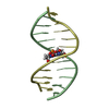
 Movie
Movie Controller


+ Open data

- Basic information
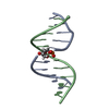
| Entry | Database: PDB / ID: 2j5y | ||||||
|---|---|---|---|---|---|---|---|
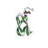
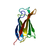
| Title | Crystal structure of the GA module from F.magna | ||||||
 Components Components | PEPTOSTREPTOCOCCAL ALBUMIN-BINDING PROTEIN | ||||||
 Keywords Keywords | PROTEIN BINDING / CELL WALL / PEPTIDOGLYCAN-ANCHOR / PROTEIN BINDING BACTERIAL ALBUMIN-BINDING THREE-HELIX BUNDLE | ||||||
| Function / homology |  Function and homology information Function and homology informationAlbumin-binding domain / Repeat of unknown function DUF5633 / Family of unknown function (DUF5633) / GA-like domain / GA-like domain / Immunoglobulin/albumin-binding domain superfamily / LPXTG cell wall anchor motif / Gram-positive cocci surface proteins LPxTG motif profile. / LPXTG cell wall anchor domain / Helicase, Ruva Protein; domain 3 ...Albumin-binding domain / Repeat of unknown function DUF5633 / Family of unknown function (DUF5633) / GA-like domain / GA-like domain / Immunoglobulin/albumin-binding domain superfamily / LPXTG cell wall anchor motif / Gram-positive cocci surface proteins LPxTG motif profile. / LPXTG cell wall anchor domain / Helicase, Ruva Protein; domain 3 / Orthogonal Bundle / Mainly Alpha Similarity search - Domain/homology | ||||||
| Biological species |  PEPTOSTREPTOCOCCUS MAGNUS (bacteria) PEPTOSTREPTOCOCCUS MAGNUS (bacteria) | ||||||
| Method |  X-RAY DIFFRACTION / SYNCHROTRON / MOLECULAR REPLACEMENT / Resolution: 1.4 Å X-RAY DIFFRACTION / SYNCHROTRON / MOLECULAR REPLACEMENT / Resolution: 1.4 Å | ||||||
 Authors Authors | Lejon, S. / Cramer, J.F. / Nordberg, P.A. / Lundqvist, T. / Valegard, K. | ||||||
 Citation Citation | Journal: FEBS Lett. / Year: 2007 Title: Crystal Structure of a Bacterial Albumin-Binding Domain at 1.4A Resolution. Authors: Cramer, J.F. / Nordberg, P.A. / Hajdu, J. / Lejon, S. #1: Journal: J.Biol.Chem. / Year: 2004Title: Crystal Structure and Biological Implications of a Bacterial Albumin-Binding Module in Complex with Human Serum Albumin Authors: Lejon, S. / Frick, I.-M. / Bjorck, L. / Wikstrom, M. / Svensson, S. | ||||||
| History |
|
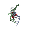
- Structure visualization
| Structure viewer | Molecule: MolmilJmol/JSmol |
|---|
- Downloads & links
-Download
| PDBx/mmCIF format | 2j5y.cif.gz | 71.5 KB | Display | PDBx/mmCIF format |
|---|---|---|---|---|
| PDB format | pdb2j5y.ent.gz | 53.3 KB | Display | PDB format |
| PDBx/mmJSON format | 2j5y.json.gz | Tree view | PDBx/mmJSON format | |
| Others |  Other downloads Other downloads |
-Validation report
| Arichive directory | https://data.pdbj.org/pub/pdb/validation_reports/j5/2j5yftp://data.pdbj.org/pub/pdb/validation_reports/j5/2j5y | HTTPS FTP |
|---|
-Related structure data
| Related structure data |  1tf0S S: Starting model for refinement |
|---|---|
| Similar structure data |


-Links
 PDBj
PDBj- Assembly
| Deposited unit | 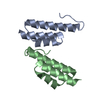
| ||||||||||||||||||
|---|---|---|---|---|---|---|---|---|---|---|---|---|---|---|---|---|---|---|---|
| 1 | 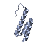
| ||||||||||||||||||
| 2 | 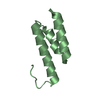
| ||||||||||||||||||
| Unit cell |
| ||||||||||||||||||
| Noncrystallographic symmetry (NCS) | NCS domain:
NCS domain segments: Component-ID: 1 / Ens-ID: 1 / Beg auth comp-ID: LEU / Beg label comp-ID: LEU / End auth comp-ID: ALA / End label comp-ID: ALA / Refine code: 2 / Auth seq-ID: -7 - 53 / Label seq-ID: 1 - 61
NCS oper: (Code: given Matrix: (-1, 0.00041, -0.0006), Vector: |
-Components
| #1: Protein | Mass: 6819.855 Da / Num. of mol.: 2 / Fragment: ALBUMIN-BINDING DOMAIN, RESIDUES 213-265 Source method: isolated from a genetically manipulated source Source: (gene. exp.) PEPTOSTREPTOCOCCUS MAGNUS (bacteria) / Plasmid: PET28 / Production host: #2: Water | ChemComp-HOH / |  Mass: 18.015 Da / Num. of mol.: 183 / Source method: isolated from a natural source / Formula: H2O Mass: 18.015 Da / Num. of mol.: 183 / Source method: isolated from a natural source / Formula: H2OSequence details | GA DOMAIN OF PROTEIN PAB, SPANNING AMINO ACIDS 213-265. | |
|---|
-Experimental details
-Experiment
| Experiment | Method: X-RAY DIFFRACTION / Number of used crystals: 1 |
|---|
- Sample preparation
| Crystal | Density Matthews: 1.88 Å3/Da / Density % sol: 34.5 % |
|---|---|
| Crystal grow | pH: 7.5 Details: 22-26% (W/V) PEG 3350, 50 MM POTASSIUM PHOSPHATE PH 7.5, 100 MM AMMONIUM ACETATE |
-Data collection
| Diffraction | Mean temperature: 100 K |
|---|---|
| Diffraction source | Source: SYNCHROTRON / Site: ESRF  / Beamline: ID23-2 / Wavelength: 0.873 / Beamline: ID23-2 / Wavelength: 0.873 |
| Detector | Type: MARRESEARCH / Detector: CCD / Date: Dec 9, 2005 / Details: PT-COATED MIRRORS IN KB GEOMETRY |
| Radiation | Monochromator: SI(111) / Protocol: SINGLE WAVELENGTH / Monochromatic (M) / Laue (L): M / Scattering type: x-ray |
| Radiation wavelength | Wavelength: 0.873 Å / Relative weight: 1 |
| Reflection | Resolution: 1.4→40 Å / Num. obs: 22273 / % possible obs: 94.4 % / Observed criterion σ(I): 2 / Redundancy: 2.5 % / Rmerge(I) obs: 0.07 / Net I/σ(I): 11.7 |
| Reflection shell | Resolution: 1.4→1.48 Å / Redundancy: 2.2 % / Rmerge(I) obs: 0.24 / Mean I/σ(I) obs: 2.8 / % possible all: 72.6 |
- Processing
| Software |
| ||||||||||||||||||||||||||||||||||||||||||||||||||||||||||||||||||||||||||||||||||||||||||||||||||||||||||||||||||||||||||||||||||||||||||||||||||||||||||||||||||||||||||||||||||||||
|---|---|---|---|---|---|---|---|---|---|---|---|---|---|---|---|---|---|---|---|---|---|---|---|---|---|---|---|---|---|---|---|---|---|---|---|---|---|---|---|---|---|---|---|---|---|---|---|---|---|---|---|---|---|---|---|---|---|---|---|---|---|---|---|---|---|---|---|---|---|---|---|---|---|---|---|---|---|---|---|---|---|---|---|---|---|---|---|---|---|---|---|---|---|---|---|---|---|---|---|---|---|---|---|---|---|---|---|---|---|---|---|---|---|---|---|---|---|---|---|---|---|---|---|---|---|---|---|---|---|---|---|---|---|---|---|---|---|---|---|---|---|---|---|---|---|---|---|---|---|---|---|---|---|---|---|---|---|---|---|---|---|---|---|---|---|---|---|---|---|---|---|---|---|---|---|---|---|---|---|---|---|---|---|
| Refinement | Method to determine structure: MOLECULAR REPLACEMENT Starting model: PDB ENTRY 1TF0, CHAIN B Resolution: 1.4→40.56 Å / Cor.coef. Fo:Fc: 0.966 / Cor.coef. Fo:Fc free: 0.947 / SU B: 2.055 / SU ML: 0.038 / Cross valid method: THROUGHOUT / ESU R: 0.074 / ESU R Free: 0.069 / Stereochemistry target values: MAXIMUM LIKELIHOOD Details: HYDROGENS HAVE BEEN ADDED IN THE RIDING POSITIONS.DISORDERED SIDE CHAINS HAVE BEEN MODELLED WITH ZERO OCCUPANCY.
| ||||||||||||||||||||||||||||||||||||||||||||||||||||||||||||||||||||||||||||||||||||||||||||||||||||||||||||||||||||||||||||||||||||||||||||||||||||||||||||||||||||||||||||||||||||||
| Solvent computation | Ion probe radii: 0.8 Å / Shrinkage radii: 0.8 Å / VDW probe radii: 1.4 Å / Solvent model: MASK | ||||||||||||||||||||||||||||||||||||||||||||||||||||||||||||||||||||||||||||||||||||||||||||||||||||||||||||||||||||||||||||||||||||||||||||||||||||||||||||||||||||||||||||||||||||||
| Displacement parameters | Biso mean: 14.77 Å2
| ||||||||||||||||||||||||||||||||||||||||||||||||||||||||||||||||||||||||||||||||||||||||||||||||||||||||||||||||||||||||||||||||||||||||||||||||||||||||||||||||||||||||||||||||||||||
| Refinement step | Cycle: LAST / Resolution: 1.4→40.56 Å
| ||||||||||||||||||||||||||||||||||||||||||||||||||||||||||||||||||||||||||||||||||||||||||||||||||||||||||||||||||||||||||||||||||||||||||||||||||||||||||||||||||||||||||||||||||||||
| Refine LS restraints |
|