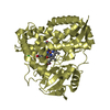
- PDB-4r7a: Crystal Structure of RBBP4 bound to PHF6 peptide -
+
Open data
ID or keywords:
Loading...
-
Basic information
Entry
Database: PDB / ID: 4r7a
Title
Crystal Structure of RBBP4 bound to PHF6 peptide
Components
Histone-binding protein RBBP4
PHD finger protein 6
Keywords
GENE REGULATION / WD40 repeat domain / nuclear
Function / homology
Function and homology information
CAF-1 complex / NURF complex / NuRD complex / regulation of cell fate specification / negative regulation of stem cell population maintenance / DNA replication-dependent chromatin assembly / ESC/E(Z) complex / Transcription of E2F targets under negative control by p107 (RBL1) and p130 (RBL2) in complex with HDAC1 / regulation of stem cell differentiation / Polo-like kinase mediated events ...CAF-1 complex / NURF complex / NuRD complex / regulation of cell fate specification / negative regulation of stem cell population maintenance / DNA replication-dependent chromatin assembly / ESC/E(Z) complex / Transcription of E2F targets under negative control by p107 (RBL1) and p130 (RBL2) in complex with HDAC1 / regulation of stem cell differentiation / Polo-like kinase mediated events / Transcription of E2F targets under negative control by DREAM complex / ATPase complex / G1/S-Specific Transcription / Sin3-type complex / histone deacetylase complex / positive regulation of stem cell population maintenance / Transcriptional Regulation by E2F6 / RNA Polymerase I Transcription Initiation / G0 and Early G1 / ribonucleoprotein complex binding / Cyclin E associated events during G1/S transition / Transcriptional regulation of brown and beige adipocyte differentiation by EBF2 / Cyclin A:Cdk2-associated events at S phase entry / Regulation of TP53 Activity through Acetylation / Deposition of new CENPA-containing nucleosomes at the centromere / negative regulation of cell migration / Regulation of PTEN gene transcription / ERCC6 (CSB) and EHMT2 (G9a) positively regulate rRNA expression / PRC2 methylates histones and DNA / Regulation of endogenous retroelements by KRAB-ZFP proteins / Defective pyroptosis / HDACs deacetylate histones / Regulation of endogenous retroelements by Piwi-interacting RNAs (piRNAs) / negative regulation of transforming growth factor beta receptor signaling pathway / phosphoprotein binding / tubulin binding / brain development / Negative Regulation of CDH1 Gene Transcription / kinetochore / PKMTs methylate histone lysines / Activation of anterior HOX genes in hindbrain development during early embryogenesis / histone deacetylase binding / HCMV Early Events / nucleosome assembly / scaffold protein binding / histone binding / Oxidative Stress Induced Senescence / Potential therapeutics for SARS / DNA replication / chromosome, telomeric region / RNA polymerase II cis-regulatory region sequence-specific DNA binding / chromatin remodeling / negative regulation of cell population proliferation / DNA repair / negative regulation of DNA-templated transcription / regulation of transcription by RNA polymerase II / regulation of DNA-templated transcription / positive regulation of DNA-templated transcription / chromatin / DNA-templated transcription / nucleolus / enzyme binding / negative regulation of transcription by RNA polymerase II / protein-containing complex / DNA binding / RNA binding / zinc ion binding / nucleoplasm / nucleus / cytosol Similarity search - Function
In the structure databanks used in Yorodumi, some data are registered as the other names, "COVID-19 virus" and "2019-nCoV". Here are the details of the virus and the list of structure data.
Jan 31, 2019. EMDB accession codes are about to change! (news from PDBe EMDB page)
EMDB accession codes are about to change! (news from PDBe EMDB page)
The allocation of 4 digits for EMDB accession codes will soon come to an end. Whilst these codes will remain in use, new EMDB accession codes will include an additional digit and will expand incrementally as the available range of codes is exhausted. The current 4-digit format prefixed with “EMD-” (i.e. EMD-XXXX) will advance to a 5-digit format (i.e. EMD-XXXXX), and so on. It is currently estimated that the 4-digit codes will be depleted around Spring 2019, at which point the 5-digit format will come into force.
The EM Navigator/Yorodumi systems omit the EMD- prefix.
Related info.:Q: What is EMD? / ID/Accession-code notation in Yorodumi/EM Navigator
Yorodumi is a browser for structure data from EMDB, PDB, SASBDB, etc.
This page is also the successor to EM Navigator detail page, and also detail information page/front-end page for Omokage search.
The word "yorodu" (or yorozu) is an old Japanese word meaning "ten thousand". "mi" (miru) is to see.
Related info.:EMDB / PDB / SASBDB / Comparison of 3 databanks / Yorodumi Search / Aug 31, 2016. New EM Navigator & Yorodumi / Yorodumi Papers / Jmol/JSmol / Function and homology information / Changes in new EM Navigator and Yorodumi
 Movie
Movie Controller


 Open data
 Basic information
 Components
 Keywords
 Function and homology information
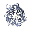
 Homo sapiens (human)
 X-RAY DIFFRACTION /
  Authors
 Citation
 Structure visualization
 Downloads & links
 Other downloads
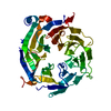


 PDBj
PDBj


 Assembly

 Spodoptera frugiperda (fall armyworm) / References: UniProt: Q09028
 Mass: 92.094 Da / Num. of mol.: 1 / Source method: obtained synthetically / Formula: C3H8O3
 Mass: 18.015 Da / Num. of mol.: 267 / Source method: isolated from a natural source / Formula: H2O
 Sample preparation
 / Beamline: BL17U / Wavelength: 0.979 Å
 Processing