- PDB-4qxk: Joint X-ray/neutron structure of PKGIbeta in complex with cGMP -
+
Open data
ID or keywords:
Loading...
-
Basic information
Entry
Database: PDB / ID: 4qxk
Title
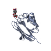
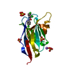
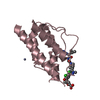
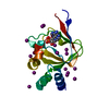
Joint X-ray/neutron structure of PKGIbeta in complex with cGMP
Components
cGMP-dependent protein kinase 1
Keywords
SIGNALING PROTEIN / cyclic GMP (cGMP) / Type I cGMP Dependent Protein Kinase (PKG I) / Cyclic Nucleotide Selectivity / Serine/threonine Protein Kinase / Signal Transduction / Second Messengers / Hydrogen Bonding / Solvent Accessibility
Function / homology
Function and homology information
negative regulation of inositol phosphate biosynthetic process / negative regulation of glutamate secretion / cGMP-dependent protein kinase / cGMP-dependent protein kinase activity / bone growth / cell growth involved in cardiac muscle cell development / regulation of testosterone biosynthetic process / collateral sprouting / negative regulation of platelet aggregation / positive regulation of circadian rhythm ...negative regulation of inositol phosphate biosynthetic process / negative regulation of glutamate secretion / cGMP-dependent protein kinase / cGMP-dependent protein kinase activity / bone growth / cell growth involved in cardiac muscle cell development / regulation of testosterone biosynthetic process / collateral sprouting / negative regulation of platelet aggregation / positive regulation of circadian rhythm / relaxation of vascular associated smooth muscle / Rap1 signalling / regulation of vascular permeability / : / mitogen-activated protein kinase p38 binding / cGMP effects / negative regulation of vascular associated smooth muscle cell migration / forebrain development / dendrite development / cGMP binding / negative regulation of vascular associated smooth muscle cell proliferation / spermatid development / cerebellum development / acrosomal vesicle / calcium channel regulator activity / sarcolemma / neuron migration / positive regulation of cytosolic calcium ion concentration / Ca2+ pathway / protein kinase activity / protein serine kinase activity / Golgi apparatus / signal transduction / nucleoplasm / ATP binding / identical protein binding / plasma membrane / cytoplasm / cytosol Similarity search - Function
cGMP-dependent protein kinase, N-terminal coiled-coil domain / Coiled-coil N-terminus of cGMP-dependent protein kinase / cGMP-dependent kinase / cGMP-dependent protein kinase, catalytic domain / Cyclic nucleotide-binding domain signature 2. / Cyclic nucleotide-binding domain signature 1. / Cyclic nucleotide-binding, conserved site / Cyclic nucleotide-monophosphate binding domain / Cyclic nucleotide-binding domain / cAMP/cGMP binding motif profile. ...cGMP-dependent protein kinase, N-terminal coiled-coil domain / Coiled-coil N-terminus of cGMP-dependent protein kinase / cGMP-dependent kinase / cGMP-dependent protein kinase, catalytic domain / Cyclic nucleotide-binding domain signature 2. / Cyclic nucleotide-binding domain signature 1. / Cyclic nucleotide-binding, conserved site / Cyclic nucleotide-monophosphate binding domain / Cyclic nucleotide-binding domain / cAMP/cGMP binding motif profile. / Cyclic nucleotide-binding domain / Cyclic nucleotide-binding domain superfamily / Jelly Rolls / Extension to Ser/Thr-type protein kinases / AGC-kinase, C-terminal / AGC-kinase C-terminal domain profile. / RmlC-like jelly roll fold / Jelly Rolls / Serine/threonine-protein kinase, active site / Serine/Threonine protein kinases active-site signature. / Protein kinase domain / Serine/Threonine protein kinases, catalytic domain / Protein kinase, ATP binding site / Protein kinases ATP-binding region signature. / Protein kinase domain profile. / Protein kinase domain / Protein kinase-like domain superfamily / Sandwich / Mainly Beta Similarity search - Domain/homology
Mass: 18.015 Da / Num. of mol.: 111 / Source method: isolated from a natural source / Formula: D2O
-
Experimental details
-
Experiment
Experiment
Method
Number of used crystals
X-RAY DIFFRACTION
1
NEUTRON DIFFRACTION
1
-
Sample preparation
Crystal
Density Matthews: 1.84 Å3/Da / Density % sol: 33.08 %
Crystal grow
Temperature: 293 K / Method: vapor diffusion, sitting drop / pH: 6.5 Details: 1.4 M Na3Citrate, pH 6.5 and 0.2 M NaI solutions, VAPOR DIFFUSION, SITTING DROP, temperature 293K
In the structure databanks used in Yorodumi, some data are registered as the other names, "COVID-19 virus" and "2019-nCoV". Here are the details of the virus and the list of structure data.
Jan 31, 2019. EMDB accession codes are about to change! (news from PDBe EMDB page)
EMDB accession codes are about to change! (news from PDBe EMDB page)
The allocation of 4 digits for EMDB accession codes will soon come to an end. Whilst these codes will remain in use, new EMDB accession codes will include an additional digit and will expand incrementally as the available range of codes is exhausted. The current 4-digit format prefixed with “EMD-” (i.e. EMD-XXXX) will advance to a 5-digit format (i.e. EMD-XXXXX), and so on. It is currently estimated that the 4-digit codes will be depleted around Spring 2019, at which point the 5-digit format will come into force.
The EM Navigator/Yorodumi systems omit the EMD- prefix.
Related info.:Q: What is EMD? / ID/Accession-code notation in Yorodumi/EM Navigator
Yorodumi is a browser for structure data from EMDB, PDB, SASBDB, etc.
This page is also the successor to EM Navigator detail page, and also detail information page/front-end page for Omokage search.
The word "yorodu" (or yorozu) is an old Japanese word meaning "ten thousand". "mi" (miru) is to see.
Related info.:EMDB / PDB / SASBDB / Comparison of 3 databanks / Yorodumi Search / Aug 31, 2016. New EM Navigator & Yorodumi / Yorodumi Papers / Jmol/JSmol / Function and homology information / Changes in new EM Navigator and Yorodumi
 Movie
Movie Controller


 Open data
 Basic information
 Components
 Keywords
 Function and homology information
 Homo sapiens (human)
 X-RAY DIFFRACTION / NEUTRON DIFFRACTION / NUCLEAR REACTOR /
  Authors
 Citation
 Structure visualization
 Downloads & links
 Other downloads


 PDBj
PDBj


 Assembly


 Mass: 345.205 Da / Num. of mol.: 1 / Source method: obtained synthetically / Formula: C10H12N5O7P
 Mass: 22.990 Da / Num. of mol.: 1 / Source method: obtained synthetically / Formula: Na
 Mass: 18.015 Da / Num. of mol.: 111 / Source method: isolated from a natural source / Formula: D2O
 Sample preparation
 Processing