 Movie
Movie Controller

[English] 日本語
 Yorodumi
- PDB-4quk: Crystal Structure of Cinnamyl-Alcohol Dehydrogenase 2 Mutant K169A -
+ Open data

- Basic information
| Entry | Database: PDB / ID: 4quk | ||||||
|---|---|---|---|---|---|---|---|
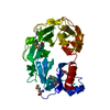
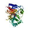
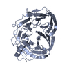
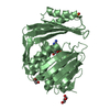
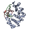
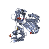
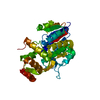
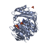
| Title | Crystal Structure of Cinnamyl-Alcohol Dehydrogenase 2 Mutant K169A | ||||||
 Components Components | Dihydroflavonol-4-reductase | ||||||
 Keywords Keywords | OXIDOREDUCTASE / dehydrogenase / monolignol / mutant / short-chain dehydrogenase/reductase / Rossmann fold | ||||||
| Function / homology |  Function and homology information Function and homology informationcinnamoyl-CoA reductase / cinnamoyl-CoA reductase activity / phenylpropanoid biosynthetic process / oxidoreductase activity, acting on the CH-OH group of donors, NAD or NADP as acceptor / cytoplasm Similarity search - Function | ||||||
| Biological species |  | ||||||
| Method |  X-RAY DIFFRACTION / FOURIER SYNTHESIS / Resolution: 1.9 Å X-RAY DIFFRACTION / FOURIER SYNTHESIS / Resolution: 1.9 Å | ||||||
 Authors Authors | Pan, H. / Wang, X. | ||||||
 Citation Citation | Journal: Plant Cell / Year: 2014 Title: Structural Studies of Cinnamoyl-CoA Reductase and Cinnamyl-Alcohol Dehydrogenase, Key Enzymes of Monolignol Biosynthesis. Authors: Pan, H. / Zhou, R. / Louie, G.V. / Muhlemann, J.K. / Bomati, E.K. / Bowman, M.E. / Dudareva, N. / Dixon, R.A. / Noel, J.P. / Wang, X. | ||||||
| History |
|
- Structure visualization
| Structure viewer | Molecule: MolmilJmol/JSmol |
|---|
- Downloads & links
-Download
| PDBx/mmCIF format | 4quk.cif.gz | 81.4 KB | Display | PDBx/mmCIF format |
|---|---|---|---|---|
| PDB format | pdb4quk.ent.gz | 59 KB | Display | PDB format |
| PDBx/mmJSON format | 4quk.json.gz | Tree view | PDBx/mmJSON format | |
| Others |  Other downloads Other downloads |
-Validation report
| Arichive directory | https://data.pdbj.org/pub/pdb/validation_reports/qu/4qukftp://data.pdbj.org/pub/pdb/validation_reports/qu/4quk | HTTPS FTP |
|---|
-Related structure data
| Related structure data |  4qtzSC  4r1sC  4r1tC  4r1uC S: Starting model for refinement C: citing same article ( |
|---|---|
| Similar structure data |


-Links
 PDBj
PDBj- Assembly
| Deposited unit | 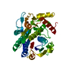
| ||||||||
|---|---|---|---|---|---|---|---|---|---|
| 1 |
| ||||||||
| Unit cell |
|
-Components
| #1: Protein | Mass: 35105.203 Da / Num. of mol.: 1 / Mutation: K169A Source method: isolated from a genetically manipulated source Source: (gene. exp.)  References: UniProt: G7IYC1, Oxidoreductases; Acting on the CH-OH group of donors; With NAD+ or NADP+ as acceptor |
|---|---|
| #2: Water | ChemComp-HOH /  Mass: 18.015 Da / Num. of mol.: 359 / Source method: isolated from a natural source / Formula: H2O Mass: 18.015 Da / Num. of mol.: 359 / Source method: isolated from a natural source / Formula: H2O |
-Experimental details
-Experiment
| Experiment | Method: X-RAY DIFFRACTION / Number of used crystals: 1 |
|---|
- Sample preparation
| Crystal | Density Matthews: 2.19 Å3/Da / Density % sol: 43.72 % |
|---|---|
| Crystal grow | Temperature: 277 K / Method: vapor diffusion / pH: 6.5 Details: 25% PEG3350, 0.1 M Bis-Tris, pH 6.5, VAPOR DIFFUSION, temperature 277K |
-Data collection
| Diffraction | Mean temperature: 93 K |
|---|---|
| Diffraction source | Source: ROTATING ANODE / Type: RIGAKU RUH3R / Wavelength: 1.5418 Å |
| Detector | Type: RIGAKU RAXIS IV++ / Detector: IMAGE PLATE / Date: Aug 16, 2012 |
| Radiation | Protocol: SINGLE WAVELENGTH / Monochromatic (M) / Laue (L): M / Scattering type: x-ray |
| Radiation wavelength | Wavelength: 1.5418 Å / Relative weight: 1 |
| Reflection | Resolution: 1.9→28.56 Å / Num. all: 24741 / Num. obs: 24741 / % possible obs: 98.6 % / Observed criterion σ(I): -3 / Biso Wilson estimate: 15.3 Å2 / Rmerge(I) obs: 0.039 / Net I/σ(I): 42.2 |
| Reflection shell | Resolution: 1.9→2.02 Å / Rmerge(I) obs: 0.13 / Mean I/σ(I) obs: 10.2 / % possible all: 88.7 |
- Processing
| Software |
| ||||||||||||||||||||||||||||||||||||||||||||||||||||||||||||
|---|---|---|---|---|---|---|---|---|---|---|---|---|---|---|---|---|---|---|---|---|---|---|---|---|---|---|---|---|---|---|---|---|---|---|---|---|---|---|---|---|---|---|---|---|---|---|---|---|---|---|---|---|---|---|---|---|---|---|---|---|---|
| Refinement | Method to determine structure: FOURIER SYNTHESIS Starting model: PDB ENTRY 4QTZ Resolution: 1.9→28.56 Å / Rfactor Rfree error: 0.005 / Data cutoff high absF: 221965.15 / Data cutoff low absF: 0 / Isotropic thermal model: RESTRAINED / Cross valid method: THROUGHOUT / σ(F): 0 / Stereochemistry target values: Engh & Huber / Details: BULK SOLVENT MODEL USED
| ||||||||||||||||||||||||||||||||||||||||||||||||||||||||||||
| Solvent computation | Solvent model: FLAT MODEL / Bsol: 61.291 Å2 / ksol: 0.4 e/Å3 | ||||||||||||||||||||||||||||||||||||||||||||||||||||||||||||
| Displacement parameters | Biso mean: 20.6 Å2
| ||||||||||||||||||||||||||||||||||||||||||||||||||||||||||||
| Refine analyze |
| ||||||||||||||||||||||||||||||||||||||||||||||||||||||||||||
| Refinement step | Cycle: LAST / Resolution: 1.9→28.56 Å
| ||||||||||||||||||||||||||||||||||||||||||||||||||||||||||||
| Refine LS restraints |
| ||||||||||||||||||||||||||||||||||||||||||||||||||||||||||||
| LS refinement shell | Resolution: 1.9→2.02 Å / Rfactor Rfree error: 0.013 / Total num. of bins used: 6
| ||||||||||||||||||||||||||||||||||||||||||||||||||||||||||||
| Xplor file |
|
