 Movie
Movie Controller

[English] 日本語
 Yorodumi
- PDB-4qt7: Crystal structure of the c-Src SH3 domain in complex with a pepti... -
+ Open data

- Basic information
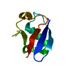
| Entry | Database: PDB / ID: 4qt7 | ||||||
|---|---|---|---|---|---|---|---|
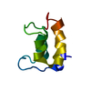
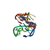
| Title | Crystal structure of the c-Src SH3 domain in complex with a peptide from the Hepatitis C virus NS5A-protein | ||||||
 Components Components |
| ||||||
 Keywords Keywords | TRANSFERASE/TRANSFERASE ACTIVATOR / beta shandwich / SH3 domain / Proline rich motifs / VIRAL PROTEIN / TRANSFERASE-TRANSFERASE ACTIVATOR complex | ||||||
| Function / homology |  Function and homology information Function and homology informationSignaling by ERBB2 / Nuclear signaling by ERBB4 / Signaling by SCF-KIT / Regulation of KIT signaling / Signaling by EGFR / GAB1 signalosome / Regulation of gap junction activity / FCGR activation / PECAM1 interactions / Co-stimulation by CD28 ...Signaling by ERBB2 / Nuclear signaling by ERBB4 / Signaling by SCF-KIT / Regulation of KIT signaling / Signaling by EGFR / GAB1 signalosome / Regulation of gap junction activity / FCGR activation / PECAM1 interactions / Co-stimulation by CD28 / Co-inhibition by CTLA4 / EPHA-mediated growth cone collapse / Ephrin signaling / G alpha (i) signalling events / GP1b-IX-V activation signalling / Thrombin signalling through proteinase activated receptors (PARs) / VEGFR2 mediated cell proliferation / RET signaling / Receptor Mediated Mitophagy / ADP signalling through P2Y purinoceptor 1 / RAF activation / PIP3 activates AKT signaling / EPH-ephrin mediated repulsion of cells / PI5P, PP2A and IER3 Regulate PI3K/AKT Signaling / Activated NTRK3 signals through PI3K / Downstream signal transduction / Downregulation of ERBB4 signaling / Cyclin D associated events in G1 / Regulation of RUNX3 expression and activity / MAP2K and MAPK activation / Integrin signaling / GRB2:SOS provides linkage to MAPK signaling for Integrins / DCC mediated attractive signaling / MET activates PTK2 signaling / Extra-nuclear estrogen signaling / EPHB-mediated forward signaling / p130Cas linkage to MAPK signaling for integrins / VEGFA-VEGFR2 Pathway / connexin binding / negative regulation of intrinsic apoptotic signaling pathway / progesterone receptor signaling pathway / immune system process / negative regulation of extrinsic apoptotic signaling pathway / non-membrane spanning protein tyrosine kinase activity / non-specific protein-tyrosine kinase / ribonucleoside triphosphate phosphatase activity / epidermal growth factor receptor signaling pathway / cell-cell junction / cell junction / protein tyrosine kinase activity / protein phosphatase binding / cell differentiation / cytoskeleton / regulation of cell cycle / cell adhesion / endosome membrane / mitochondrial inner membrane / signaling receptor binding / serine-type endopeptidase activity / cysteine-type endopeptidase activity / focal adhesion / RNA-directed RNA polymerase activity / heme binding / perinuclear region of cytoplasm / protein-containing complex / zinc ion binding / ATP binding / nucleus / membrane / plasma membrane / cytosol Similarity search - Function | ||||||
| Biological species |   Hepatitis C virus Hepatitis C virus | ||||||
| Method |  X-RAY DIFFRACTION / SYNCHROTRON / MOLECULAR REPLACEMENT / molecular replacement / Resolution: 1.55 Å X-RAY DIFFRACTION / SYNCHROTRON / MOLECULAR REPLACEMENT / molecular replacement / Resolution: 1.55 Å | ||||||
 Authors Authors | Camara-Artigas, A. / Bacarizo, J. | ||||||
 Citation Citation | Journal: J.Struct.Biol. / Year: 2015 Title: Structure of the c-Src-SH3 domain in complex with a proline-rich motif of NS5A protein from the hepatitis C virus. Authors: Bacarizo, J. / Martinez-Rodriguez, S. / Camara-Artigas, A. | ||||||
| History |
|
- Structure visualization
| Structure viewer | Molecule: MolmilJmol/JSmol |
|---|
- Downloads & links
-Download
| PDBx/mmCIF format | 4qt7.cif.gz | 50.7 KB | Display | PDBx/mmCIF format |
|---|---|---|---|---|
| PDB format | pdb4qt7.ent.gz | 35.4 KB | Display | PDB format |
| PDBx/mmJSON format | 4qt7.json.gz | Tree view | PDBx/mmJSON format | |
| Others |  Other downloads Other downloads |
-Validation report
| Arichive directory | https://data.pdbj.org/pub/pdb/validation_reports/qt/4qt7ftp://data.pdbj.org/pub/pdb/validation_reports/qt/4qt7 | HTTPS FTP |
|---|
-Related structure data
| Related structure data |  4jz4S S: Starting model for refinement |
|---|---|
| Similar structure data |


-Links
 PDBj
PDBj
- Assembly
| Deposited unit | 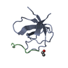
| ||||||||
|---|---|---|---|---|---|---|---|---|---|
| 1 |
| ||||||||
| Unit cell |
|
-Components
| #1: Protein | Mass: 6905.498 Da / Num. of mol.: 1 / Fragment: SH3 domain, UNP residues 85-141 Source method: isolated from a genetically manipulated source Source: (gene. exp.)  References: UniProt: P00523, non-specific protein-tyrosine kinase |
|---|---|
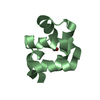
| #2: Protein/peptide | Mass: 1314.624 Da / Num. of mol.: 1 / Fragment: Proline rich peptide, UNP residues 349-359 / Source method: obtained synthetically / Details: The peptide was chemically synthesized. / Source: (synth.) Hepatitis C virus / References: UniProt: V5RF47 |
| #3: Water | ChemComp-HOH /  Mass: 18.015 Da / Num. of mol.: 94 / Source method: isolated from a natural source / Formula: H2O Mass: 18.015 Da / Num. of mol.: 94 / Source method: isolated from a natural source / Formula: H2O |
| Has protein modification | Y |
-Experimental details
-Experiment
| Experiment | Method: X-RAY DIFFRACTION / Number of used crystals: 1 |
|---|
- Sample preparation
| Crystal | Density Matthews: 1.61 Å3/Da / Density % sol: 23.59 % Description: THE ENTRY CONTAINS FRIEDEL PAIRS IN F_PLUS/MINUS COLUMNS Mosaicity: 0.23 ° |
|---|---|
| Crystal grow | Temperature: 298 K / Method: vapor diffusion, hanging drop / pH: 7 Details: 2M Ammonium sulphate, 0.1M sodium chloride, 0.86mM methyl-beta-cyclodextrin, 0.1M Hepes, pH 7, vapor diffusion, hanging drop, temperature 298K |
-Data collection
| Diffraction | Mean temperature: 100 K | ||||||||||||||||||||||||
|---|---|---|---|---|---|---|---|---|---|---|---|---|---|---|---|---|---|---|---|---|---|---|---|---|---|
| Diffraction source | Source: SYNCHROTRON / Site: ESRF  / Beamline: ID14-1 / Wavelength: 0.9334 Å / Beamline: ID14-1 / Wavelength: 0.9334 Å | ||||||||||||||||||||||||
| Detector | Detector: CCD / Date: Nov 28, 2012 Details: vertical focusing mirror (VFM) and a horizontal focusing mirror (HFM), manufactured by IRELEC. | ||||||||||||||||||||||||
| Radiation | Monochromator: Si(111) channel-cut crystal monochromator and a pair of KB mirrors Protocol: SINGLE WAVELENGTH / Monochromatic (M) / Laue (L): M / Scattering type: x-ray | ||||||||||||||||||||||||
| Radiation wavelength | Wavelength: 0.9334 Å / Relative weight: 1 | ||||||||||||||||||||||||
| Reflection | Resolution: 1.55→24.98 Å / Num. obs: 14570 / % possible obs: 98.2 % / Observed criterion σ(F): 0 / Observed criterion σ(I): 0 / Redundancy: 9.6 % / Biso Wilson estimate: 7.42 Å2 / Rmerge(I) obs: 0.091 / Net I/σ(I): 23.7 / Scaling rejects: 6 | ||||||||||||||||||||||||
| Reflection shell | Diffraction-ID: 1 / Rejects: _
|
-Phasing
| Phasing | Method: molecular replacement |
|---|
- Processing
| Software |
| ||||||||||||||||||||||||||||||||||||||||||
|---|---|---|---|---|---|---|---|---|---|---|---|---|---|---|---|---|---|---|---|---|---|---|---|---|---|---|---|---|---|---|---|---|---|---|---|---|---|---|---|---|---|---|---|
| Refinement | Method to determine structure: MOLECULAR REPLACEMENT Starting model: 4JZ4 Resolution: 1.55→24.979 Å / SU ML: 0.17 / Cross valid method: THROUGHOUT / σ(F): 0.01 / Phase error: 22.26 / Stereochemistry target values: ML Details: THE ENTRY CONTAINS FRIEDEL PAIRS IN F_PLUS/MINUS COLUMNS
| ||||||||||||||||||||||||||||||||||||||||||
| Solvent computation | Shrinkage radii: 0.9 Å / VDW probe radii: 1.11 Å / Solvent model: FLAT BULK SOLVENT MODEL | ||||||||||||||||||||||||||||||||||||||||||
| Displacement parameters | Biso max: 42.62 Å2 / Biso mean: 10.5396 Å2 / Biso min: 3 Å2 | ||||||||||||||||||||||||||||||||||||||||||
| Refinement step | Cycle: LAST / Resolution: 1.55→24.979 Å
| ||||||||||||||||||||||||||||||||||||||||||
| Refine LS restraints |
| ||||||||||||||||||||||||||||||||||||||||||
| LS refinement shell | Refine-ID: X-RAY DIFFRACTION / Total num. of bins used: 5
|
