- PDB-4qmj: The XMAP215 family drives microtubule polymerization using a stru... -
+
Open data
ID or keywords:
Loading...
-
Basic information
Entry
Database: PDB / ID: 4qmj
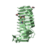
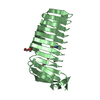
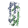
Title
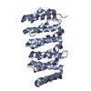
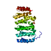
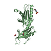
The XMAP215 family drives microtubule polymerization using a structurally diverse TOG array
Components
Cytoskeleton-associated protein 5
Keywords
PROTEIN BINDING / TOG DOMAIN
Function / homology
Function and homology information
microtubule plus end polymerase activity / establishment or maintenance of microtubule cytoskeleton polarity / RNA transport / positive regulation of microtubule nucleation / microtubule plus-end binding / microtubule depolymerization / spindle organization / centrosome cycle / centrosome duplication / microtubule polymerization ...microtubule plus end polymerase activity / establishment or maintenance of microtubule cytoskeleton polarity / RNA transport / positive regulation of microtubule nucleation / microtubule plus-end binding / microtubule depolymerization / spindle organization / centrosome cycle / centrosome duplication / microtubule polymerization / ribonucleoprotein complex binding / Amplification of signal from unattached kinetochores via a MAD2 inhibitory signal / Loss of Nlp from mitotic centrosomes / Loss of proteins required for interphase microtubule organization from the centrosome / Recruitment of mitotic centrosome proteins and complexes / Recruitment of NuMA to mitotic centrosomes / Anchoring of the basal body to the plasma membrane / Mitotic Prometaphase / EML4 and NUDC in mitotic spindle formation / AURKA Activation by TPX2 / Resolution of Sister Chromatid Cohesion / mitotic spindle organization / RHO GTPases Activate Formins / kinetochore / spindle pole / Separation of Sister Chromatids / Regulation of PLK1 Activity at G2/M Transition / microtubule binding / cadherin binding / cell division / centrosome / protein-containing complex / membrane / cytoplasm / cytosol Similarity search - Function
CLASP N-terminal domain / XMAP215 family / CLASP N terminal / : / XMAP215/Dis1/CLASP, TOG domain / TOG domain / TOG / HEAT repeat profile. / HEAT, type 2 / Leucine-rich Repeat Variant ...CLASP N-terminal domain / XMAP215 family / CLASP N terminal / : / XMAP215/Dis1/CLASP, TOG domain / TOG domain / TOG / HEAT repeat profile. / HEAT, type 2 / Leucine-rich Repeat Variant / Leucine-rich Repeat Variant / Alpha Horseshoe / Armadillo-like helical / Armadillo-type fold / Mainly Alpha Similarity search - Domain/homology
Mass: 18.015 Da / Num. of mol.: 48 / Source method: isolated from a natural source / Formula: H2O
Has protein modification
Y
-
Experimental details
-
Experiment
Experiment
Method: X-RAY DIFFRACTION / Number of used crystals: 1
-
Sample preparation
Crystal
Density Matthews: 1.98 Å3/Da / Density % sol: 37.8 %
Crystal grow
Temperature: 293.15 K / Method: vapor diffusion, hanging drop / pH: 7.5 Details: 10 mg/ml protein was added to an equal volume of a mother liquor containing 27.5% PEG 4000, 100 mM Tris pH 7.5, and 70 mM MgCl2, VAPOR DIFFUSION, HANGING DROP, temperature 293.15K
In the structure databanks used in Yorodumi, some data are registered as the other names, "COVID-19 virus" and "2019-nCoV". Here are the details of the virus and the list of structure data.
Jan 31, 2019. EMDB accession codes are about to change! (news from PDBe EMDB page)
EMDB accession codes are about to change! (news from PDBe EMDB page)
The allocation of 4 digits for EMDB accession codes will soon come to an end. Whilst these codes will remain in use, new EMDB accession codes will include an additional digit and will expand incrementally as the available range of codes is exhausted. The current 4-digit format prefixed with “EMD-” (i.e. EMD-XXXX) will advance to a 5-digit format (i.e. EMD-XXXXX), and so on. It is currently estimated that the 4-digit codes will be depleted around Spring 2019, at which point the 5-digit format will come into force.
The EM Navigator/Yorodumi systems omit the EMD- prefix.
Related info.:Q: What is EMD? / ID/Accession-code notation in Yorodumi/EM Navigator
Yorodumi is a browser for structure data from EMDB, PDB, SASBDB, etc.
This page is also the successor to EM Navigator detail page, and also detail information page/front-end page for Omokage search.
The word "yorodu" (or yorozu) is an old Japanese word meaning "ten thousand". "mi" (miru) is to see.
Related info.:EMDB / PDB / SASBDB / Comparison of 3 databanks / Yorodumi Search / Aug 31, 2016. New EM Navigator & Yorodumi / Yorodumi Papers / Jmol/JSmol / Function and homology information / Changes in new EM Navigator and Yorodumi
 Movie
Movie Controller

 Yorodumi
 Open data
 Basic information
 Components
 Keywords
 Function and homology information
 Homo sapiens (human)
 X-RAY DIFFRACTION /
  Authors
 Citation
 Structure visualization
 Downloads & links
 Other downloads


 PDBj
PDBj


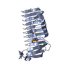
 Assembly
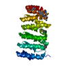

 Mass: 18.015 Da / Num. of mol.: 48 / Source method: isolated from a natural source / Formula: H2O
 Sample preparation
 / Beamline: 22-ID / Wavelength: 0.97926 Å
 Processing