 Movie
Movie Controller

[English] 日本語
 Yorodumi
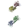
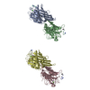
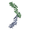
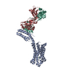
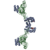
- PDB-4ql0: Crystal Structure Analysis of the Membrane Transporter FhaC (doub... -
+ Open data

- Basic information
| Entry | Database: PDB / ID: 4ql0 | ||||||
|---|---|---|---|---|---|---|---|
| Title | Crystal Structure Analysis of the Membrane Transporter FhaC (double mutant V169T, I176N) | ||||||
 Components Components | Filamentous hemagglutinin transporter protein FhaC | ||||||
 Keywords Keywords | PROTEIN TRANSPORT / Beta-barrel / POTRA domain / Outer Membrane | ||||||
| Function / homology |  Function and homology information Function and homology informationtype V protein secretion system complex / protein secretion by the type V secretion system / porin activity / pore complex / transmembrane protein transporter activity / monoatomic ion transport / cell outer membrane Similarity search - Function | ||||||
| Biological species |  Bordetella pertussis (bacteria) Bordetella pertussis (bacteria) | ||||||
| Method |  X-RAY DIFFRACTION / SYNCHROTRON / FOURIER SYNTHESIS / Resolution: 2.5 Å X-RAY DIFFRACTION / SYNCHROTRON / FOURIER SYNTHESIS / Resolution: 2.5 Å | ||||||
 Authors Authors | Maier, T. / Clantin, B. / Gruss, F. / Dewitte, F. / Delattre, A.S. / Jacob-Dubuisson, F. / Hiller, S. / Villeret, V. | ||||||
 Citation Citation | Journal: Nat Commun / Year: 2015 Title: Conserved Omp85 lid-lock structure and substrate recognition in FhaC Authors: Maier, T. / Clantin, B. / Gruss, F. / Dewitte, F. / Delattre, A.S. / Jacob-Dubuisson, F. / Hiller, S. / Villeret, V. #1: Journal: Science / Year: 2007 Title: Structure of the membrane protein FhaC: a member of the Omp85-TpsB transporter superfamily Authors: Clantin, B. / Delattre, A.S. / Rucktooa, P. / Saint, N. / Meli, A.C. / Locht, C. / Jacob-Dubuisson, F. / Villeret, V. #2: Journal: Mol.Microbiol. / Year: 2011 Title: Substrate recognition by the POTRA domains of TpsB transporter FhaC Authors: Delattre, A.S. / Saint, N. / Clantin, B. / Willery, E. / Lippens, G. / Locht, C. / Villeret, V. / Jacob-Dubuisson, F. | ||||||
| History |
|
- Structure visualization
| Structure viewer | Molecule: MolmilJmol/JSmol |
|---|
- Downloads & links
-Download
| PDBx/mmCIF format | 4ql0.cif.gz | 420.9 KB | Display | PDBx/mmCIF format |
|---|---|---|---|---|
| PDB format | pdb4ql0.ent.gz | 351.7 KB | Display | PDB format |
| PDBx/mmJSON format | 4ql0.json.gz | Tree view | PDBx/mmJSON format | |
| Others |  Other downloads Other downloads |
-Validation report
| Arichive directory | https://data.pdbj.org/pub/pdb/validation_reports/ql/4ql0ftp://data.pdbj.org/pub/pdb/validation_reports/ql/4ql0 | HTTPS FTP |
|---|
-Related structure data
| Related structure data |  4qkySC S: Starting model for refinement C: citing same article ( |
|---|---|
| Similar structure data |


-Links
 PDBj
PDBj- Assembly
| Deposited unit | 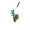
| ||||||||
|---|---|---|---|---|---|---|---|---|---|
| 1 | 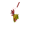
| ||||||||
| Unit cell |
| ||||||||
| Components on special symmetry positions |
|
-Components
-Protein / Sugars , 2 types, 4 molecules A
| #1: Protein | Mass: 61415.758 Da / Num. of mol.: 1 / Mutation: V169T, I176N Source method: isolated from a genetically manipulated source Source: (gene. exp.) Bordetella pertussis (bacteria) / Strain: Tohama I / ATCC BAA-589 / NCTC 13251 / Gene: BP1884, fhaC / Plasmid: pFJD138 / Production host: |
|---|---|
| #4: Sugar |  Type: D-saccharide / Mass: 292.369 Da / Num. of mol.: 3 Type: D-saccharide / Mass: 292.369 Da / Num. of mol.: 3Source method: isolated from a genetically manipulated source Formula: C14H28O6 / Comment: detergent*YM |
-Non-polymers , 7 types, 81 molecules 


| #2: Chemical | ChemComp-PGE /  Mass: 150.173 Da / Num. of mol.: 1 / Source method: obtained synthetically / Formula: C6H14O4 Mass: 150.173 Da / Num. of mol.: 1 / Source method: obtained synthetically / Formula: C6H14O4 |
|---|---|
| #3: Chemical | ChemComp-PEG /  Mass: 106.120 Da / Num. of mol.: 1 / Source method: obtained synthetically / Formula: C4H10O3 Mass: 106.120 Da / Num. of mol.: 1 / Source method: obtained synthetically / Formula: C4H10O3 |
| #5: Chemical | ChemComp-PO4 /  Mass: 94.971 Da / Num. of mol.: 1 / Source method: obtained synthetically / Formula: PO4 Mass: 94.971 Da / Num. of mol.: 1 / Source method: obtained synthetically / Formula: PO4 |
| #6: Chemical | ChemComp-1PE /  Mass: 238.278 Da / Num. of mol.: 1 / Source method: obtained synthetically / Formula: C10H22O6 / Comment: precipitant*YM Mass: 238.278 Da / Num. of mol.: 1 / Source method: obtained synthetically / Formula: C10H22O6 / Comment: precipitant*YM |
| #7: Chemical | ChemComp-PG4 /  Mass: 194.226 Da / Num. of mol.: 1 / Source method: obtained synthetically / Formula: C8H18O5 / Comment: precipitant*YM Mass: 194.226 Da / Num. of mol.: 1 / Source method: obtained synthetically / Formula: C8H18O5 / Comment: precipitant*YM |
| #8: Chemical | ChemComp-P6G /  Mass: 282.331 Da / Num. of mol.: 1 / Source method: obtained synthetically / Formula: C12H26O7 / Comment: precipitant*YM Mass: 282.331 Da / Num. of mol.: 1 / Source method: obtained synthetically / Formula: C12H26O7 / Comment: precipitant*YM |
| #9: Water | ChemComp-HOH / Mass: 18.015 Da / Num. of mol.: 75 / Source method: isolated from a natural source / Formula: H2O |
-Experimental details
-Experiment
| Experiment | Method: X-RAY DIFFRACTION / Number of used crystals: 1 |
|---|
- Sample preparation
| Crystal | Density Matthews: 3.29 Å3/Da / Density % sol: 62.62 % |
|---|---|
| Crystal grow | Temperature: 298 K / Method: vapor diffusion, hanging drop / pH: 6.5 Details: 34% PEG 1000, 1% beta-octyl-glucoside, 500mM imidazole, 26mg/ml protein, pH 6.5, VAPOR DIFFUSION, HANGING DROP, temperature 298K |
-Data collection
| Diffraction | Mean temperature: 100 K |
|---|---|
| Diffraction source | Source: SYNCHROTRON / Site: ESRF  / Beamline: ID14-4 / Wavelength: 0.93928 Å / Beamline: ID14-4 / Wavelength: 0.93928 Å |
| Detector | Type: ADSC QUANTUM 315r / Detector: CCD / Date: Nov 10, 2010 |
| Radiation | Monochromator: channel cut ESRF monochromator / Protocol: SINGLE WAVELENGTH / Monochromatic (M) / Laue (L): M / Scattering type: x-ray |
| Radiation wavelength | Wavelength: 0.93928 Å / Relative weight: 1 |
| Reflection | Resolution: 2.5→41.95 Å / Num. all: 28332 / Num. obs: 28332 / % possible obs: 99.69 % / Observed criterion σ(F): 0 / Biso Wilson estimate: 74.17 Å2 |
| Reflection shell | Resolution: 2.5→2.65 Å / % possible all: 98.8 |
- Processing
| Software |
| |||||||||||||||||||||||||||||||||||||||||||||||||||||||||||||||||||||||||||||||||||||||||||||||||||||||||||||||||||||||||||||
|---|---|---|---|---|---|---|---|---|---|---|---|---|---|---|---|---|---|---|---|---|---|---|---|---|---|---|---|---|---|---|---|---|---|---|---|---|---|---|---|---|---|---|---|---|---|---|---|---|---|---|---|---|---|---|---|---|---|---|---|---|---|---|---|---|---|---|---|---|---|---|---|---|---|---|---|---|---|---|---|---|---|---|---|---|---|---|---|---|---|---|---|---|---|---|---|---|---|---|---|---|---|---|---|---|---|---|---|---|---|---|---|---|---|---|---|---|---|---|---|---|---|---|---|---|---|---|
| Refinement | Method to determine structure: FOURIER SYNTHESIS Starting model: 4QKY Resolution: 2.5→41.95 Å / Cor.coef. Fo:Fc: 0.9141 / Cor.coef. Fo:Fc free: 0.8894 / Cross valid method: THROUGHOUT / σ(F): 0 / SU R Blow DPI: 0.326 / SU Rfree Blow DPI: 0.246 / Stereochemistry target values: Engh & Huber / Details: isomorphous replacement using pdb entry 4QKY
| |||||||||||||||||||||||||||||||||||||||||||||||||||||||||||||||||||||||||||||||||||||||||||||||||||||||||||||||||||||||||||||
| Displacement parameters | Biso mean: 78.93 Å2
| |||||||||||||||||||||||||||||||||||||||||||||||||||||||||||||||||||||||||||||||||||||||||||||||||||||||||||||||||||||||||||||
| Refine analyze | Luzzati coordinate error obs: 0.503 Å | |||||||||||||||||||||||||||||||||||||||||||||||||||||||||||||||||||||||||||||||||||||||||||||||||||||||||||||||||||||||||||||
| Refinement step | Cycle: LAST / Resolution: 2.5→41.95 Å
| |||||||||||||||||||||||||||||||||||||||||||||||||||||||||||||||||||||||||||||||||||||||||||||||||||||||||||||||||||||||||||||
| Refine LS restraints |
| |||||||||||||||||||||||||||||||||||||||||||||||||||||||||||||||||||||||||||||||||||||||||||||||||||||||||||||||||||||||||||||
| LS refinement shell | Resolution: 2.5→2.59 Å / Total num. of bins used: 14
| |||||||||||||||||||||||||||||||||||||||||||||||||||||||||||||||||||||||||||||||||||||||||||||||||||||||||||||||||||||||||||||
| Refinement TLS params. | Method: refined / Refine-ID: X-RAY DIFFRACTION
| |||||||||||||||||||||||||||||||||||||||||||||||||||||||||||||||||||||||||||||||||||||||||||||||||||||||||||||||||||||||||||||
| Refinement TLS group |
|
