 Movie
Movie Controller

[English] 日本語
 Yorodumi
- PDB-4qg3: Crystal structure of mutant ribosomal protein G219V TthL1 in comp... -
+ Open data

- Basic information
| Entry | Database: PDB / ID: 4qg3 | ||||||
|---|---|---|---|---|---|---|---|
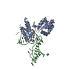
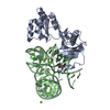
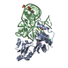
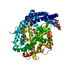
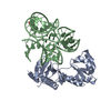
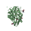
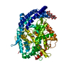
| Title | Crystal structure of mutant ribosomal protein G219V TthL1 in complex with 80nt 23S RNA from Thermus thermophilus | ||||||
 Components Components |
| ||||||
 Keywords Keywords | RIBOSOMAL PROTEIN/RNA / Rossmann Fold / RIBOSOMAL PROTEIN / rRNA / rRNA BINDING / RIBOSOME / RIBOSOMAL PROTEIN-RNA complex | ||||||
| Function / homology |  Function and homology information Function and homology informationregulation of translation / large ribosomal subunit / tRNA binding / rRNA binding / structural constituent of ribosome / translation Similarity search - Function | ||||||
| Biological species |   Thermus thermophilus (bacteria) Thermus thermophilus (bacteria) | ||||||
| Method |  X-RAY DIFFRACTION / MOLECULAR REPLACEMENT / Resolution: 2 Å X-RAY DIFFRACTION / MOLECULAR REPLACEMENT / Resolution: 2 Å | ||||||
 Authors Authors | Gabdulkhakov, A.G. / Nevskaya, N.A. / Nikonov, S.V. | ||||||
 Citation Citation | Journal: Acta Crystallogr.,Sect.D / Year: 2015 Title: Protein-RNA affinity of ribosomal protein L1 mutants does not correlate with the number of intermolecular interactions. Authors: Tishchenko, S. / Kostareva, O. / Gabdulkhakov, A. / Mikhaylina, A. / Nikonova, E. / Nevskaya, N. / Sarskikh, A. / Piendl, W. / Garber, M. / Nikonov, S. | ||||||
| History |
|
- Structure visualization
| Structure viewer | Molecule: MolmilJmol/JSmol |
|---|
- Downloads & links
-Download
| PDBx/mmCIF format | 4qg3.cif.gz | 113.3 KB | Display | PDBx/mmCIF format |
|---|---|---|---|---|
| PDB format | pdb4qg3.ent.gz | 80.2 KB | Display | PDB format |
| PDBx/mmJSON format | 4qg3.json.gz | Tree view | PDBx/mmJSON format | |
| Others |  Other downloads Other downloads |
-Validation report
| Arichive directory | https://data.pdbj.org/pub/pdb/validation_reports/qg/4qg3ftp://data.pdbj.org/pub/pdb/validation_reports/qg/4qg3 | HTTPS FTP |
|---|
-Related structure data
| Related structure data | 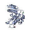 4qgbC 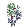 4qviC 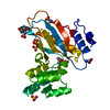 4reoC 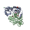 3u4mS S: Starting model for refinement C: citing same article ( |
|---|---|
| Similar structure data |


-Links
 PDBj
PDBj


- Assembly
| Deposited unit | 
| ||||||||
|---|---|---|---|---|---|---|---|---|---|
| 1 |
| ||||||||
| Unit cell |
|
-Components
-Protein / RNA chain , 2 types, 2 molecules AB
| #1: Protein | Mass: 24778.582 Da / Num. of mol.: 1 / Mutation: G219V Source method: isolated from a genetically manipulated source Source: (gene. exp.) Thermus thermophilus (bacteria) / Gene: rplA, rpl1 / Plasmid: pET11a-PL/TthL1 G219V / Production host: |
|---|---|
| #2: RNA chain | Mass: 25974.475 Da / Num. of mol.: 1 / Fragment: 80 nt fragment of 23S rRNA Source method: isolated from a genetically manipulated source Source: (gene. exp.) Thermus thermophilus (bacteria) / Plasmid: pUC18/rRNATth / Production host: |
-Non-polymers , 5 types, 279 molecules 


| #3: Chemical | ChemComp-SO4 /  Mass: 96.063 Da / Num. of mol.: 5 / Source method: obtained synthetically / Formula: SO4 Mass: 96.063 Da / Num. of mol.: 5 / Source method: obtained synthetically / Formula: SO4#4: Chemical | ChemComp-IPA /  Mass: 60.095 Da / Num. of mol.: 8 / Source method: obtained synthetically / Formula: C3H8O / Comment: alkaloid*YM Mass: 60.095 Da / Num. of mol.: 8 / Source method: obtained synthetically / Formula: C3H8O / Comment: alkaloid*YM#5: Chemical | ChemComp-BME /  Mass: 78.133 Da / Num. of mol.: 4 / Source method: obtained synthetically / Formula: C2H6OS Mass: 78.133 Da / Num. of mol.: 4 / Source method: obtained synthetically / Formula: C2H6OS#6: Chemical | ChemComp-URE / |  Mass: 60.055 Da / Num. of mol.: 1 / Source method: obtained synthetically / Formula: CH4N2O Mass: 60.055 Da / Num. of mol.: 1 / Source method: obtained synthetically / Formula: CH4N2O#7: Water | ChemComp-HOH / | Mass: 18.015 Da / Num. of mol.: 261 / Source method: isolated from a natural source / Formula: H2O |
|---|
-Experimental details
-Experiment
| Experiment | Method: X-RAY DIFFRACTION / Number of used crystals: 1 |
|---|
- Sample preparation
| Crystal | Density Matthews: 2.36 Å3/Da / Density % sol: 47.99 % |
|---|---|
| Crystal grow | Temperature: 295 K / Method: vapor diffusion, hanging drop / pH: 8.5 Details: 1.8 M Ammonium sulfate, 0.05 M Bis-Tris,pH 8.5, VAPOR DIFFUSION, HANGING DROP, temperature 295K |
-Data collection
| Diffraction | Mean temperature: 110 K |
|---|---|
| Diffraction source | Source: ROTATING ANODE / Type: BRUKER AXS MICROSTAR / Wavelength: 1.5418 Å |
| Detector | Type: Bruker Platinum 135 / Detector: CCD / Date: Mar 1, 2013 |
| Radiation | Monochromator: Montel 200 / Protocol: SINGLE WAVELENGTH / Monochromatic (M) / Laue (L): M / Scattering type: x-ray |
| Radiation wavelength | Wavelength: 1.5418 Å / Relative weight: 1 |
| Reflection | Resolution: 2→50 Å / Num. all: 33273 / Num. obs: 33152 / % possible obs: 99.6 % / Observed criterion σ(F): 0 / Observed criterion σ(I): 0 |
| Reflection shell | Resolution: 2→2.1 Å / % possible all: 99.3 |
- Processing
| Software |
| ||||||||||||||||||||||||||||||||||||||||||||||||||||||||||||||||||||||||||||||||||||
|---|---|---|---|---|---|---|---|---|---|---|---|---|---|---|---|---|---|---|---|---|---|---|---|---|---|---|---|---|---|---|---|---|---|---|---|---|---|---|---|---|---|---|---|---|---|---|---|---|---|---|---|---|---|---|---|---|---|---|---|---|---|---|---|---|---|---|---|---|---|---|---|---|---|---|---|---|---|---|---|---|---|---|---|---|---|
| Refinement | Method to determine structure: MOLECULAR REPLACEMENT Starting model: PDB ENTRY 3U4M Resolution: 2→22.981 Å / SU ML: 0.24 / σ(F): 1.35 / Phase error: 22.32 / Stereochemistry target values: ML
| ||||||||||||||||||||||||||||||||||||||||||||||||||||||||||||||||||||||||||||||||||||
| Solvent computation | Shrinkage radii: 0.9 Å / VDW probe radii: 1.11 Å / Solvent model: FLAT BULK SOLVENT MODEL | ||||||||||||||||||||||||||||||||||||||||||||||||||||||||||||||||||||||||||||||||||||
| Refinement step | Cycle: LAST / Resolution: 2→22.981 Å
| ||||||||||||||||||||||||||||||||||||||||||||||||||||||||||||||||||||||||||||||||||||
| Refine LS restraints |
| ||||||||||||||||||||||||||||||||||||||||||||||||||||||||||||||||||||||||||||||||||||
| LS refinement shell |
|
