 Movie
Movie Controller

[English] 日本語
 Yorodumi
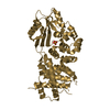
- PDB-4qdk: Crystal structure of magnesium protoporphyrin IX methyltransferas... -
+ Open data

- Basic information
| Entry | Database: PDB / ID: 4qdk | ||||||
|---|---|---|---|---|---|---|---|
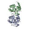
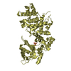
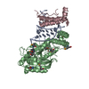
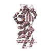
| Title | Crystal structure of magnesium protoporphyrin IX methyltransferase (ChlM) from Synechocystis PCC 6803 with bound SAH | ||||||
 Components Components | Magnesium-protoporphyrin O-methyltransferase | ||||||
 Keywords Keywords | TRANSFERASE / Methyltransferase / Magnesium protoporphyrin IX / S-adenosyl Homocysteine | ||||||
| Function / homology |  Function and homology information Function and homology informationmagnesium protoporphyrin IX methyltransferase / magnesium protoporphyrin IX methyltransferase activity / light-independent chlorophyll biosynthetic process / photosynthesis / methyltransferase activity / methylation Similarity search - Function | ||||||
| Biological species |  | ||||||
| Method |  X-RAY DIFFRACTION / SYNCHROTRON / MOLECULAR REPLACEMENT / Resolution: 1.7 Å X-RAY DIFFRACTION / SYNCHROTRON / MOLECULAR REPLACEMENT / Resolution: 1.7 Å | ||||||
 Authors Authors | Chen, X. / Wang, X. / Liu, L. | ||||||
 Citation Citation | Journal: J.Biol.Chem. / Year: 2014 Title: Structural insights into the catalytic mechanism of Synechocystis magnesium protoporphyrin IX O-methyltransferase (ChlM). Authors: Chen, X. / Wang, X. / Feng, J. / Chen, Y. / Fang, Y. / Zhao, S. / Zhao, A. / Zhang, M. / Liu, L. | ||||||
| History |
|
- Structure visualization
| Structure viewer | Molecule: MolmilJmol/JSmol |
|---|
- Downloads & links
-Download
| PDBx/mmCIF format | 4qdk.cif.gz | 198.3 KB | Display | PDBx/mmCIF format |
|---|---|---|---|---|
| PDB format | pdb4qdk.ent.gz | 157.2 KB | Display | PDB format |
| PDBx/mmJSON format | 4qdk.json.gz | Tree view | PDBx/mmJSON format | |
| Others |  Other downloads Other downloads |
-Validation report
| Summary document | 4qdk_validation.pdf.gz | 1 MB | Display | wwPDB validaton report |
|---|---|---|---|---|
| Full document | 4qdk_full_validation.pdf.gz | 1 MB | Display | |
| Data in XML | 4qdk_validation.xml.gz | 22 KB | Display | |
| Data in CIF | 4qdk_validation.cif.gz | 31.7 KB | Display | |
| Arichive directory | https://data.pdbj.org/pub/pdb/validation_reports/qd/4qdkftp://data.pdbj.org/pub/pdb/validation_reports/qd/4qdk | HTTPS FTP |
-Related structure data
| Related structure data |  4qdjSC S: Starting model for refinement C: citing same article ( |
|---|---|
| Similar structure data |


-Links
 PDBj
PDBj- Assembly
| Deposited unit | 
| ||||||||
|---|---|---|---|---|---|---|---|---|---|
| 1 |
| ||||||||
| 2 | 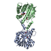
| ||||||||
| Unit cell |
|
-Components
| #1: Protein | Mass: 26179.801 Da / Num. of mol.: 2 Source method: isolated from a genetically manipulated source Source: (gene. exp.) References: UniProt: Q55467, magnesium protoporphyrin IX methyltransferase #2: Chemical |   Type: L-peptide linking / Mass: 384.411 Da / Num. of mol.: 2 / Source method: obtained synthetically / Formula: C14H20N6O5S Type: L-peptide linking / Mass: 384.411 Da / Num. of mol.: 2 / Source method: obtained synthetically / Formula: C14H20N6O5S#3: Chemical |   Mass: 92.094 Da / Num. of mol.: 2 / Source method: obtained synthetically / Formula: C3H8O3 Mass: 92.094 Da / Num. of mol.: 2 / Source method: obtained synthetically / Formula: C3H8O3#4: Water | ChemComp-HOH / |  Mass: 18.015 Da / Num. of mol.: 372 / Source method: isolated from a natural source / Formula: H2O Mass: 18.015 Da / Num. of mol.: 372 / Source method: isolated from a natural source / Formula: H2O |
|---|
-Experimental details
-Experiment
| Experiment | Method: X-RAY DIFFRACTION / Number of used crystals: 1 |
|---|
- Sample preparation
| Crystal | Density Matthews: 2.44 Å3/Da / Density % sol: 49.58 % |
|---|---|
| Crystal grow | Temperature: 277 K / Method: vapor diffusion, sitting drop / pH: 5.8 Details: 0.1M citrate sodium pH 5.8, 10% PEG 4000, 20% isopropanol, VAPOR DIFFUSION, SITTING DROP, temperature 277K |
-Data collection
| Diffraction | Mean temperature: 100 K |
|---|---|
| Diffraction source | Source: SYNCHROTRON / Site: SSRF  / Beamline: BL17U / Wavelength: 0.9793 Å / Beamline: BL17U / Wavelength: 0.9793 Å |
| Detector | Type: ADSC QUANTUM 315r / Detector: CCD / Date: Jul 24, 2013 |
| Radiation | Monochromator: double crystal / Protocol: SINGLE WAVELENGTH / Monochromatic (M) / Laue (L): M / Scattering type: x-ray |
| Radiation wavelength | Wavelength: 0.9793 Å / Relative weight: 1 |
| Reflection | Resolution: 1.7→50 Å / Num. all: 54547 / Num. obs: 54275 / % possible obs: 99.5 % / Observed criterion σ(F): 2 / Observed criterion σ(I): 2 |
| Reflection shell | Resolution: 1.7→1.76 Å / % possible all: 98.9 |
- Processing
| Software |
| ||||||||||||||||||||||||||||||||||||||||||||||||||||||||||||||||||||||||||||||||||||||||||||||||||||||||||||||||||
|---|---|---|---|---|---|---|---|---|---|---|---|---|---|---|---|---|---|---|---|---|---|---|---|---|---|---|---|---|---|---|---|---|---|---|---|---|---|---|---|---|---|---|---|---|---|---|---|---|---|---|---|---|---|---|---|---|---|---|---|---|---|---|---|---|---|---|---|---|---|---|---|---|---|---|---|---|---|---|---|---|---|---|---|---|---|---|---|---|---|---|---|---|---|---|---|---|---|---|---|---|---|---|---|---|---|---|---|---|---|---|---|---|---|---|---|
| Refinement | Method to determine structure: MOLECULAR REPLACEMENT Starting model: 4QDJ Resolution: 1.7→36.043 Å / SU ML: 0.17 / σ(F): 1.38 / Phase error: 23.27 / Stereochemistry target values: ML
| ||||||||||||||||||||||||||||||||||||||||||||||||||||||||||||||||||||||||||||||||||||||||||||||||||||||||||||||||||
| Solvent computation | Shrinkage radii: 0.9 Å / VDW probe radii: 1.11 Å / Solvent model: FLAT BULK SOLVENT MODEL | ||||||||||||||||||||||||||||||||||||||||||||||||||||||||||||||||||||||||||||||||||||||||||||||||||||||||||||||||||
| Refinement step | Cycle: LAST / Resolution: 1.7→36.043 Å
| ||||||||||||||||||||||||||||||||||||||||||||||||||||||||||||||||||||||||||||||||||||||||||||||||||||||||||||||||||
| Refine LS restraints |
| ||||||||||||||||||||||||||||||||||||||||||||||||||||||||||||||||||||||||||||||||||||||||||||||||||||||||||||||||||
| LS refinement shell | Refine-ID: X-RAY DIFFRACTION / Total num. of bins used: 18
|
