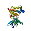
[English] 日本語
Yorodumi
- PDB-4q9s: Crystal Structure of human Focal Adhesion Kinase (Fak) bound to C... -

+
Open data


ID or keywords:

Loading...

-
Basic information

Entry
Database: PDB / ID: 4q9s
TitleCrystal Structure of human Focal Adhesion Kinase (Fak) bound to Compound1 (3,5-DIHYDRO[1,2,4]TRIAZINO[3,4-C][1,4]BENZOXAZIN-2(1H)-ONE)
ComponentsFocal adhesion kinase 1
KeywordsTransferase/transferase inhibitor / Kinase catalytic domain / Transferase-transferase inhibitor complex
Function / homology
Function and homology information


netrin-activated signaling pathway / regulation of substrate adhesion-dependent cell spreading / regulation of endothelial cell migration / regulation of epithelial cell migration / detection of muscle stretch / JUN kinase binding / signal complex assembly / positive regulation of ubiquitin-dependent protein catabolic process / positive regulation of macrophage proliferation / DCC mediated attractive signaling ...netrin-activated signaling pathway / regulation of substrate adhesion-dependent cell spreading / regulation of endothelial cell migration / regulation of epithelial cell migration / detection of muscle stretch / JUN kinase binding / signal complex assembly / positive regulation of ubiquitin-dependent protein catabolic process / positive regulation of macrophage proliferation / DCC mediated attractive signaling / positive regulation of fibroblast migration / Signal regulatory protein family interactions / regulation of GTPase activity / growth hormone receptor signaling pathway / MET activates PTK2 signaling / regulation of focal adhesion assembly / negative regulation of cell-cell adhesion / positive regulation of wound healing / p130Cas linkage to MAPK signaling for integrins / regulation of osteoblast differentiation / Apoptotic cleavage of cellular proteins / establishment of cell polarity / positive regulation of macrophage chemotaxis / regulation of cytoskeleton organization / regulation of cell adhesion mediated by integrin / Fc-gamma receptor signaling pathway involved in phagocytosis / vascular endothelial cell response to oscillatory fluid shear stress / GRB2:SOS provides linkage to MAPK signaling for Integrins / regulation of protein phosphorylation / negative regulation of anoikis / positive regulation of epithelial cell migration / ephrin receptor signaling pathway / Estrogen-dependent nuclear events downstream of ESR-membrane signaling / RHO GTPases Activate WASPs and WAVEs / positive regulation of protein kinase activity / vascular endothelial growth factor receptor signaling pathway / regulation of cell adhesion / heart morphogenesis / positive regulation of epithelial to mesenchymal transition / stress fiber / EPHB-mediated forward signaling / Integrin signaling / protein tyrosine phosphatase activity / NCAM signaling for neurite out-growth / transforming growth factor beta receptor signaling pathway / SH2 domain binding / axon guidance / molecular function activator activity / Turbulent (oscillatory, disturbed) flow shear stress activates signaling by PIEZO1 and integrins in endothelial cells / integrin-mediated signaling pathway / non-membrane spanning protein tyrosine kinase activity / FCGR3A-mediated phagocytosis / non-specific protein-tyrosine kinase / placenta development / cell motility / peptidyl-tyrosine phosphorylation / Regulation of actin dynamics for phagocytic cup formation / VEGFA-VEGFR2 Pathway / epidermal growth factor receptor signaling pathway / integrin binding / cell migration / regulation of cell shape / regulation of cell population proliferation / positive regulation of protein phosphorylation / actin binding / RAF/MAP kinase cascade / cell cortex / protein autophosphorylation / protein tyrosine kinase activity / protein phosphatase binding / angiogenesis / cytoskeleton / Extra-nuclear estrogen signaling / positive regulation of phosphatidylinositol 3-kinase/protein kinase B signal transduction / ciliary basal body / cilium / positive regulation of cell migration / focal adhesion / intracellular membrane-bounded organelle / positive regulation of cell population proliferation / centrosome / negative regulation of apoptotic process / protein kinase binding / perinuclear region of cytoplasm / ATP binding / nucleus / plasma membrane / cytosol
Similarity search - Function
Focal adhesion kinase, targeting (FAT) domain / Focal adhesion kinase, targeting (FAT) domain superfamily / Focal adhesion kinase, N-terminal / FAK1/PYK2, FERM domain C-lobe / Focal adhesion targeting region / FERM N-terminal domain / : / FAK1/PYK2, FERM domain C-lobe / FERM domain signature 2. / FERM central domain ...Focal adhesion kinase, targeting (FAT) domain / Focal adhesion kinase, targeting (FAT) domain superfamily / Focal adhesion kinase, N-terminal / FAK1/PYK2, FERM domain C-lobe / Focal adhesion targeting region / FERM N-terminal domain / : / FAK1/PYK2, FERM domain C-lobe / FERM domain signature 2. / FERM central domain / FERM/acyl-CoA-binding protein superfamily / FERM central domain / FERM superfamily, second domain / FERM domain / FERM domain profile. / Band 4.1 domain / Band 4.1 homologues / Tyrosine-protein kinase, catalytic domain / Tyrosine kinase, catalytic domain / Tyrosine protein kinases specific active-site signature. / Tyrosine-protein kinase, active site / PH-like domain superfamily / Serine-threonine/tyrosine-protein kinase, catalytic domain / Protein tyrosine and serine/threonine kinase / Ubiquitin-like domain superfamily / Phosphorylase Kinase; domain 1 / Phosphorylase Kinase; domain 1 / Transferase(Phosphotransferase) domain 1 / Transferase(Phosphotransferase); domain 1 / Protein kinase, ATP binding site / Protein kinases ATP-binding region signature. / Protein kinase domain profile. / Protein kinase domain / Protein kinase-like domain superfamily / 2-Layer Sandwich / Orthogonal Bundle / Mainly Alpha / Alpha Beta
Similarity search - Domain/homology
Chem-30G / Focal adhesion kinase 1
Similarity search - Component
Biological speciesHomo sapiens (human)
MethodX-RAY DIFFRACTION / SYNCHROTRON / MOLECULAR REPLACEMENT / Resolution: 2.07 Å
AuthorsArgiriadi, M.A. / George, D.M.
CitationJournal: J.Med.Chem. / Year: 2015
Title: Discovery of Selective and Orally Bioavailable Protein Kinase C theta (PKC theta ) Inhibitors from a Fragment Hit.
Authors: George, D.M. / Breinlinger, E.C. / Friedman, M. / Zhang, Y. / Wang, J. / Argiriadi, M. / Bansal-Pakala, P. / Barth, M. / Duignan, D.B. / Honore, P. / Lang, Q. / Mittelstadt, S. / Potin, D. / ...Authors: George, D.M. / Breinlinger, E.C. / Friedman, M. / Zhang, Y. / Wang, J. / Argiriadi, M. / Bansal-Pakala, P. / Barth, M. / Duignan, D.B. / Honore, P. / Lang, Q. / Mittelstadt, S. / Potin, D. / Rundell, L. / Edmunds, J.J.
History
DepositionMay 1, 2014Deposition site: RCSB / Processing site: RCSB
Revision 1.0Jul 2, 2014Provider: repository / Type: Initial release
Revision 1.1Jul 30, 2014Group: Database references
Revision 1.2Sep 17, 2014Group: Source and taxonomy
Revision 1.3Sep 24, 2014Group: Source and taxonomy
Revision 1.4Jan 21, 2015Group: Database references
Revision 1.5Nov 22, 2017Group: Refinement description / Category: software
Revision 1.6Mar 6, 2024Group: Data collection / Database references ...Data collection / Database references / Derived calculations / Refinement description
Category: chem_comp_atom / chem_comp_bond ...chem_comp_atom / chem_comp_bond / database_2 / software / struct_ref_seq_dif / struct_site
Item: _database_2.pdbx_DOI / _database_2.pdbx_database_accession ..._database_2.pdbx_DOI / _database_2.pdbx_database_accession / _software.name / _struct_ref_seq_dif.details / _struct_site.pdbx_auth_asym_id / _struct_site.pdbx_auth_comp_id / _struct_site.pdbx_auth_seq_id
Revision 1.7Oct 16, 2024Group: Structure summary / Category: pdbx_entry_details / pdbx_modification_feature

-
Structure visualization

Structure viewerMolecule:
MolmilJmol/JSmol

Downloads & links

-
Assembly

Deposited unit
A: Focal adhesion kinase 1
hetero molecules


Theoretical massNumber of molelcules
Total (without water)32,3232
Polymers32,1201
Non-polymers2031
Water2,504139
1


  • Idetical with deposited unit
  • defined by author&software
TypeNameSymmetry operationNumber
identity operation1_555x,y,z1
MethodPISA
Unit cell
Length a, b, c (Å)45.840, 47.200, 63.160
Angle α, β, γ (deg.)90.00, 99.32, 90.00
Int Tables number4
Space group name H-MP1211

-
Components

#1: Protein Focal adhesion kinase 1 / FADK 1 / Focal adhesion kinase-related nonkinase / FRNK / Protein phosphatase 1 regulatory subunit ...FADK 1 / Focal adhesion kinase-related nonkinase / FRNK / Protein phosphatase 1 regulatory subunit 71 / PPP1R71 / Protein-tyrosine kinase 2 / p125FAK / pp125FAK


Mass: 32120.139 Da / Num. of mol.: 1 / Fragment: unp residues 411-686
Source method: isolated from a genetically manipulated source
Source: (gene. exp.) Homo sapiens (human) / Gene: PTK2, FAK, FAK1 / Production host: Spodoptera frugiperda (fall armyworm)
References: UniProt: Q05397, non-specific protein-tyrosine kinase
#2: Chemical ChemComp-30G / 3,5-dihydro[1,2,4]triazino[3,4-c][1,4]benzoxazin-2(1H)-one


Mass: 203.197 Da / Num. of mol.: 1 / Source method: obtained synthetically / Formula: C10H9N3O2
#3: Water ChemComp-HOH / water


Mass: 18.015 Da / Num. of mol.: 139 / Source method: isolated from a natural source / Formula: H2O
Has protein modificationY

-
Experimental details

-
Experiment

ExperimentMethod: X-RAY DIFFRACTION / Number of used crystals: 1

-
Sample preparation

CrystalDensity Matthews: 2.1 Å3/Da / Density % sol: 41.41 %
Crystal growTemperature: 290 K / Method: vapor diffusion, hanging drop / pH: 5.6
Details: Na acetate, PEG MME2000, pH 5.6, VAPOR DIFFUSION, HANGING DROP, temperature 290.0K

-
Data collection

DiffractionMean temperature: 200 K
Diffraction sourceSource: SYNCHROTRON / Site: APS / Beamline: 17-ID / Wavelength: 1 Å
DetectorType: PSI PILATUS 6M / Detector: PIXEL / Date: Aug 5, 2010
RadiationMonochromator: Si(111) / Protocol: SINGLE WAVELENGTH / Monochromatic (M) / Laue (L): M / Scattering type: x-ray
Radiation wavelengthWavelength: 1 Å / Relative weight: 1
ReflectionResolution: 2.07→47.2 Å / Num. obs: 16304 / % possible obs: 90 % / Observed criterion σ(F): 0 / Observed criterion σ(I): 0 / Biso Wilson estimate: 29.29 Å2
Reflection shell
Resolution (Å)Redundancy (%)Rmerge(I) obsMean I/σ(I) obsNum. unique allDiffraction-ID% possible all
2.065-2.0722.60.4984.8158198.8
9.559-47.22.90.02828.8181198.9

-
Processing

Software
NameVersionClassification
JDirectordata collection
PHASERphasing
BUSTER2.9.7refinement
autoPROCdata scaling
RefinementMethod to determine structure: MOLECULAR REPLACEMENT / Resolution: 2.07→18.6 Å / Cor.coef. Fo:Fc: 0.9421 / Cor.coef. Fo:Fc free: 0.9152 / SU R Cruickshank DPI: 0.199 / Cross valid method: THROUGHOUT / σ(F): 0 / Stereochemistry target values: Engh & Huber
RfactorNum. reflection% reflectionSelection details
Rfree0.2163 819 5.08 %RANDOM
Rwork0.1764 ---
all0.2163 16304 --
obs0.1784 16124 98.19 %-
Displacement parametersBiso mean: 31.94 Å2
Baniso -1Baniso -2Baniso -3
1-0.2816 Å20 Å2-5.4091 Å2
2--3.778 Å20 Å2
3----4.0595 Å2
Refine analyzeLuzzati coordinate error obs: 0.219 Å
Refinement stepCycle: LAST / Resolution: 2.07→18.6 Å
ProteinNucleic acidLigandSolventTotal
Num. atoms2024 0 15 139 2178
Refine LS restraints
Refine-IDTypeDev idealNumberRestraint functionWeight
X-RAY DIFFRACTIONt_bond_d0.012095HARMONIC2
X-RAY DIFFRACTIONt_angle_deg1.042841HARMONIC2
X-RAY DIFFRACTIONt_dihedral_angle_d732SINUSOIDAL2
X-RAY DIFFRACTIONt_incorr_chiral_ct
X-RAY DIFFRACTIONt_pseud_angle
X-RAY DIFFRACTIONt_trig_c_planes48HARMONIC2
X-RAY DIFFRACTIONt_gen_planes302HARMONIC5
X-RAY DIFFRACTIONt_it2095HARMONIC20
X-RAY DIFFRACTIONt_nbd
X-RAY DIFFRACTIONt_omega_torsion3.08
X-RAY DIFFRACTIONt_other_torsion17.13
X-RAY DIFFRACTIONt_improper_torsion
X-RAY DIFFRACTIONt_chiral_improper_torsion268SEMIHARMONIC5
X-RAY DIFFRACTIONt_sum_occupancies
X-RAY DIFFRACTIONt_utility_distance
X-RAY DIFFRACTIONt_utility_angle
X-RAY DIFFRACTIONt_utility_torsion
X-RAY DIFFRACTIONt_ideal_dist_contact2457SEMIHARMONIC4
LS refinement shellResolution: 2.07→2.21 Å / Total num. of bins used: 8
RfactorNum. reflection% reflection
Rfree0.2735 133 4.7 %
Rwork0.1917 2695 -
all0.1956 2828 -
obs--98.19 %

+
About Yorodumi

-
News

-
Feb 9, 2022. New format data for meta-information of EMDB entries

New format data for meta-information of EMDB entries

  • Version 3 of the EMDB header file is now the official format.
  • The previous official version 1.9 will be removed from the archive.

Related info.:EMDB header

External links:wwPDB to switch to version 3 of the EMDB data model

-
Aug 12, 2020. Covid-19 info

Covid-19 info

URL: https://pdbj.org/emnavi/covid19.php

New page: Covid-19 featured information page in EM Navigator.

Related info.:Covid-19 info / Mar 5, 2020. Novel coronavirus structure data

+
Mar 5, 2020. Novel coronavirus structure data

Novel coronavirus structure data

Related info.:Yorodumi Speices / Aug 12, 2020. Covid-19 info

External links:COVID-19 featured content - PDBj / Molecule of the Month (242):Coronavirus Proteases

+
Jan 31, 2019. EMDB accession codes are about to change! (news from PDBe EMDB page)

EMDB accession codes are about to change! (news from PDBe EMDB page)

  • The allocation of 4 digits for EMDB accession codes will soon come to an end. Whilst these codes will remain in use, new EMDB accession codes will include an additional digit and will expand incrementally as the available range of codes is exhausted. The current 4-digit format prefixed with “EMD-” (i.e. EMD-XXXX) will advance to a 5-digit format (i.e. EMD-XXXXX), and so on. It is currently estimated that the 4-digit codes will be depleted around Spring 2019, at which point the 5-digit format will come into force.
  • The EM Navigator/Yorodumi systems omit the EMD- prefix.

Related info.:Q: What is EMD? / ID/Accession-code notation in Yorodumi/EM Navigator

External links:EMDB Accession Codes are Changing Soon! / Contact to PDBj

+
Jul 12, 2017. Major update of PDB

Major update of PDB

  • wwPDB released updated PDB data conforming to the new PDBx/mmCIF dictionary.
  • This is a major update changing the version number from 4 to 5, and with Remediation, in which all the entries are updated.
  • In this update, many items about electron microscopy experimental information are reorganized (e.g. em_software).
  • Now, EM Navigator and Yorodumi are based on the updated data.

External links:wwPDB Remediation / Enriched Model Files Conforming to OneDep Data Standards Now Available in the PDB FTP Archive

-
Yorodumi

Thousand views of thousand structures

  • Yorodumi is a browser for structure data from EMDB, PDB, SASBDB, etc.
  • This page is also the successor to EM Navigator detail page, and also detail information page/front-end page for Omokage search.
  • The word "yorodu" (or yorozu) is an old Japanese word meaning "ten thousand". "mi" (miru) is to see.

Related info.:EMDB / PDB / SASBDB / Comparison of 3 databanks / Yorodumi Search / Aug 31, 2016. New EM Navigator & Yorodumi / Yorodumi Papers / Jmol/JSmol / Function and homology information / Changes in new EM Navigator and Yorodumi

Read more