 Movie
Movie Controller

[English] 日本語
 Yorodumi
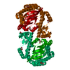
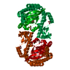
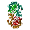
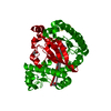
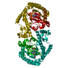
- PDB-4q8o: tRNA-Guanine Transglycosylase (TGT) Mutant V262T in Complex with ... -
+ Open data

- Basic information
| Entry | Database: PDB / ID: 4q8o | ||||||
|---|---|---|---|---|---|---|---|
| Title | tRNA-Guanine Transglycosylase (TGT) Mutant V262T in Complex with 6-Amino-2-{[2-(morpholin-4-yl)ethyl]amino}-1H,7H,8H-imidazo[4,5-g]quinazolin-8-one | ||||||
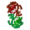
 Components Components | Queuine tRNA-ribosyltransferase | ||||||
 Keywords Keywords | Transferase/transferase inhibitor / Guanine Exchange Enzyme / preQ1 / tRNA / Transferase-transferase inhibitor complex | ||||||
| Function / homology |  Function and homology information Function and homology informationtRNA-guanosine34 preQ1 transglycosylase / tRNA-guanosine(34) queuine transglycosylase activity / tRNA queuosine(34) biosynthetic process / metal ion binding / cytosol Similarity search - Function | ||||||
| Biological species |  Zymomonas mobilis subsp. mobilis (bacteria) Zymomonas mobilis subsp. mobilis (bacteria) | ||||||
| Method |  X-RAY DIFFRACTION / SYNCHROTRON / MOLECULAR REPLACEMENT / Resolution: 1.887 Å X-RAY DIFFRACTION / SYNCHROTRON / MOLECULAR REPLACEMENT / Resolution: 1.887 Å | ||||||
 Authors Authors | Neeb, M. / Heine, A. / Klebe, G. | ||||||
 Citation Citation | Journal: To be Published Title: Creating a Resistance Model for TGT: The Effect of Mutations on Flexible lin-Benzoguanine Substituents Authors: Neeb, M. / Heine, A. / Klebe, G. | ||||||
| History |
|
- Structure visualization
| Structure viewer | Molecule: MolmilJmol/JSmol |
|---|
- Downloads & links
-Download
| PDBx/mmCIF format | 4q8o.cif.gz | 162.4 KB | Display | PDBx/mmCIF format |
|---|---|---|---|---|
| PDB format | pdb4q8o.ent.gz | 125.8 KB | Display | PDB format |
| PDBx/mmJSON format | 4q8o.json.gz | Tree view | PDBx/mmJSON format | |
| Others |  Other downloads Other downloads |
-Validation report
| Arichive directory | https://data.pdbj.org/pub/pdb/validation_reports/q8/4q8oftp://data.pdbj.org/pub/pdb/validation_reports/q8/4q8o | HTTPS FTP |
|---|
-Related structure data
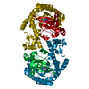
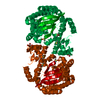
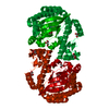
| Related structure data |  4q8mC  4q8nC  4q8pC 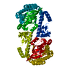 4q8qC  1pudS S: Starting model for refinement C: citing same article ( |
|---|---|
| Similar structure data |


-Links
 PDBj
PDBj- Assembly
| Deposited unit | 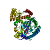
| |||||||||
|---|---|---|---|---|---|---|---|---|---|---|
| 1 | 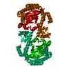
| |||||||||
| Unit cell |
| |||||||||
| Components on special symmetry positions |
|
-Components
| #1: Protein | Mass: 43071.801 Da / Num. of mol.: 1 / Mutation: V262T Source method: isolated from a genetically manipulated source Source: (gene. exp.) Zymomonas mobilis subsp. mobilis (bacteria)Strain: ATCC 31821 / ZM4 / CP4 / Gene: tgt, ZMO0363 / Plasmid: pPR-IBA2-ZM-V262T / Production host: References: UniProt: P28720, tRNA-guanosine34 preQ1 transglycosylase | ||||||
|---|---|---|---|---|---|---|---|
| #2: Chemical | ChemComp-ZN /   Mass: 65.409 Da / Num. of mol.: 1 / Source method: obtained synthetically / Formula: Zn Mass: 65.409 Da / Num. of mol.: 1 / Source method: obtained synthetically / Formula: Zn | ||||||
| #3: Chemical | ChemComp-GOL /   Mass: 92.094 Da / Num. of mol.: 4 / Source method: obtained synthetically / Formula: C3H8O3 Mass: 92.094 Da / Num. of mol.: 4 / Source method: obtained synthetically / Formula: C3H8O3#4: Chemical | ChemComp-CKR / | 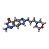  Mass: 329.357 Da / Num. of mol.: 1 / Source method: obtained synthetically / Formula: C15H19N7O2 Mass: 329.357 Da / Num. of mol.: 1 / Source method: obtained synthetically / Formula: C15H19N7O2#5: Water | ChemComp-HOH / |  Mass: 18.015 Da / Num. of mol.: 253 / Source method: isolated from a natural source / Formula: H2O Mass: 18.015 Da / Num. of mol.: 253 / Source method: isolated from a natural source / Formula: H2OSequence details | AUTHORS HAVE INDICATED THAT THERE IS A CONFLICT IN THE UNPROT SEQUENCE P28720. SEE REFERENCE: ...AUTHORS HAVE INDICATED THAT THERE IS A CONFLICT IN THE UNPROT SEQUENCE P28720. SEE REFERENCE: REUTER K.K.H., FICNER R.; J. BACTERIOL. 177:5284-5288 (1995). THE CORRECT RESIDUE IS A LYS | |
-Experimental details
-Experiment
| Experiment | Method: X-RAY DIFFRACTION / Number of used crystals: 1 |
|---|
- Sample preparation
| Crystal | Density Matthews: 2.44 Å3/Da / Density % sol: 49.53 % |
|---|---|
| Crystal grow | Temperature: 291.15 K / Method: vapor diffusion, sitting drop / pH: 5.5 Details: 100 mM MES, 10% DMSO, 13% PEG 8000, pH 5.5, VAPOR DIFFUSION, SITTING DROP, temperature 291.15K |
-Data collection
| Diffraction | Mean temperature: 100 K |
|---|---|
| Diffraction source | Source: SYNCHROTRON / Site: BESSY  / Beamline: 14.2 / Wavelength: 0.91841 Å / Beamline: 14.2 / Wavelength: 0.91841 Å |
| Detector | Type: MARMOSAIC 225 mm CCD / Detector: CCD / Date: Mar 16, 2012 / Details: Mirror |
| Radiation | Monochromator: Double Crystal Monochromator / Protocol: SINGLE WAVELENGTH / Monochromatic (M) / Laue (L): M / Scattering type: x-ray |
| Radiation wavelength | Wavelength: 0.91841 Å / Relative weight: 1 |
| Reflection | Resolution: 1.89→30 Å / Num. obs: 32679 / % possible obs: 97.9 % / Redundancy: 3.2 % / Biso Wilson estimate: 21.1 Å2 / Rsym value: 0.079 / Net I/σ(I): 15.1 |
| Reflection shell | Resolution: 1.89→1.92 Å / Redundancy: 3.2 % / Mean I/σ(I) obs: 2.9 / Num. unique all: 1625 / Rsym value: 0.434 / % possible all: 96.3 |
- Processing
| Software |
| ||||||||||||||||||||||||||||||||||||||||||||||||||||||||||||||||||||||||||||||||||||||||||||||||||||
|---|---|---|---|---|---|---|---|---|---|---|---|---|---|---|---|---|---|---|---|---|---|---|---|---|---|---|---|---|---|---|---|---|---|---|---|---|---|---|---|---|---|---|---|---|---|---|---|---|---|---|---|---|---|---|---|---|---|---|---|---|---|---|---|---|---|---|---|---|---|---|---|---|---|---|---|---|---|---|---|---|---|---|---|---|---|---|---|---|---|---|---|---|---|---|---|---|---|---|---|---|---|
| Refinement | Method to determine structure: MOLECULAR REPLACEMENT Starting model: pdb entry 1PUD Resolution: 1.887→23.631 Å / SU ML: 0.17 / Cross valid method: R-free / σ(F): 1.34 / Phase error: 19.29 / Stereochemistry target values: ML
| ||||||||||||||||||||||||||||||||||||||||||||||||||||||||||||||||||||||||||||||||||||||||||||||||||||
| Solvent computation | Shrinkage radii: 1 Å / VDW probe radii: 1.2 Å / Solvent model: FLAT BULK SOLVENT MODEL | ||||||||||||||||||||||||||||||||||||||||||||||||||||||||||||||||||||||||||||||||||||||||||||||||||||
| Refinement step | Cycle: LAST / Resolution: 1.887→23.631 Å
| ||||||||||||||||||||||||||||||||||||||||||||||||||||||||||||||||||||||||||||||||||||||||||||||||||||
| Refine LS restraints |
| ||||||||||||||||||||||||||||||||||||||||||||||||||||||||||||||||||||||||||||||||||||||||||||||||||||
| LS refinement shell |
| ||||||||||||||||||||||||||||||||||||||||||||||||||||||||||||||||||||||||||||||||||||||||||||||||||||
| Refinement TLS params. | Method: refined / Refine-ID: X-RAY DIFFRACTION
| ||||||||||||||||||||||||||||||||||||||||||||||||||||||||||||||||||||||||||||||||||||||||||||||||||||
| Refinement TLS group |
|
