 Movie
Movie Controller

[English] 日本語
 Yorodumi
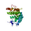
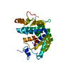
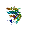
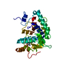
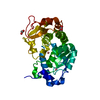
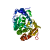
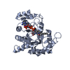
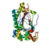
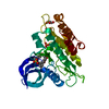
- PDB-4q3z: Crystal structure of C. violaceum phenylalanine hydroxylase D139K... -
+ Open data

- Basic information
| Entry | Database: PDB / ID: 4q3z | ||||||
|---|---|---|---|---|---|---|---|
| Title | Crystal structure of C. violaceum phenylalanine hydroxylase D139K mutation | ||||||
 Components Components | Phenylalanine-4-hydroxylase | ||||||
 Keywords Keywords | OXIDOREDUCTASE / Mutation / hydroxylase / phenylalanine hydroxylase / kinetics / metals / Chromobacterium / phenylketonuria / biopterin / Mixed alpha helix-beta sheet | ||||||
| Function / homology |  Function and homology information Function and homology informationphenylalanine 4-monooxygenase / phenylalanine 4-monooxygenase activity / L-phenylalanine catabolic process / iron ion binding Similarity search - Function | ||||||
| Biological species |  Chromobacterium violaceum (bacteria) Chromobacterium violaceum (bacteria) | ||||||
| Method |  X-RAY DIFFRACTION / SYNCHROTRON / MOLECULAR REPLACEMENT / Resolution: 1.35 Å X-RAY DIFFRACTION / SYNCHROTRON / MOLECULAR REPLACEMENT / Resolution: 1.35 Å | ||||||
 Authors Authors | Ronau, J.A. / Abu-Omar, M.M. / Das, C. | ||||||
 Citation Citation | Journal: Biochemistry / Year: 2014 Title: A conserved acidic residue in phenylalanine hydroxylase contributes to cofactor affinity and catalysis. Authors: Ronau, J.A. / Paul, L.N. / Fuchs, J.E. / Liedl, K.R. / Abu-Omar, M.M. / Das, C. | ||||||
| History |
|
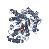
- Structure visualization
| Structure viewer | Molecule: MolmilJmol/JSmol |
|---|
- Downloads & links
-Download
| PDBx/mmCIF format | 4q3z.cif.gz | 126.1 KB | Display | PDBx/mmCIF format |
|---|---|---|---|---|
| PDB format | pdb4q3z.ent.gz | 96.9 KB | Display | PDB format |
| PDBx/mmJSON format | 4q3z.json.gz | Tree view | PDBx/mmJSON format | |
| Others |  Other downloads Other downloads |
-Validation report
| Summary document | 4q3z_validation.pdf.gz | 425.2 KB | Display | wwPDB validaton report |
|---|---|---|---|---|
| Full document | 4q3z_full_validation.pdf.gz | 426.3 KB | Display | |
| Data in XML | 4q3z_validation.xml.gz | 12.8 KB | Display | |
| Data in CIF | 4q3z_validation.cif.gz | 18 KB | Display | |
| Arichive directory | https://data.pdbj.org/pub/pdb/validation_reports/q3/4q3zftp://data.pdbj.org/pub/pdb/validation_reports/q3/4q3z | HTTPS FTP |
-Related structure data
| Related structure data |  4q3wC  4q3xC  4q3yC  1ltuS C: citing same article ( S: Starting model for refinement |
|---|---|
| Similar structure data |

-Links
 PDBj
PDBj

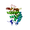
- Assembly
| Deposited unit | 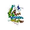
| ||||||||
|---|---|---|---|---|---|---|---|---|---|
| 1 |
| ||||||||
| Unit cell |
|
-Components
| #1: Protein | Mass: 33661.109 Da / Num. of mol.: 1 / Mutation: D139K Source method: isolated from a genetically manipulated source Source: (gene. exp.) Chromobacterium violaceum (bacteria)Strain: ATCC 12472 / DSM 30191 / JCM 1249 / NBRC 12614 / NCIMB 9131 / NCTC 9757 Gene: phhA, CV_3180 / Plasmid: pET3a / Production host: |
|---|---|
| #2: Chemical | ChemComp-CO /   Mass: 58.933 Da / Num. of mol.: 1 / Source method: obtained synthetically / Formula: Co Mass: 58.933 Da / Num. of mol.: 1 / Source method: obtained synthetically / Formula: Co |
| #3: Water | ChemComp-HOH /  Mass: 18.015 Da / Num. of mol.: 135 / Source method: isolated from a natural source / Formula: H2O Mass: 18.015 Da / Num. of mol.: 135 / Source method: isolated from a natural source / Formula: H2O |
-Experimental details
-Experiment
| Experiment | Method: X-RAY DIFFRACTION / Number of used crystals: 1 |
|---|
- Sample preparation
| Crystal | Density Matthews: 1.87 Å3/Da / Density % sol: 34.2 % |
|---|---|
| Crystal grow | Temperature: 293 K / Method: vapor diffusion, hanging drop / pH: 7 Details: 100 mM Na-HEPES pH 7.0, 10 mM Magnesium chloride hexahydrate, 5 mM Nickel(II) chloride hexahydrate, 15% w/v polyethylene glycol, VAPOR DIFFUSION, HANGING DROP, temperature 293K |
-Data collection
| Diffraction | Mean temperature: 100 K |
|---|---|
| Diffraction source | Source: SYNCHROTRON / Site: APS  / Beamline: 23-ID-B / Wavelength: 1.033 Å / Beamline: 23-ID-B / Wavelength: 1.033 Å |
| Detector | Type: MAR scanner 300 mm plate / Detector: IMAGE PLATE / Date: Dec 17, 2012 |
| Radiation | Monochromator: Si 111 Channel / Protocol: SINGLE WAVELENGTH / Monochromatic (M) / Laue (L): M / Scattering type: x-ray |
| Radiation wavelength | Wavelength: 1.033 Å / Relative weight: 1 |
| Reflection | Resolution: 1.35→50 Å / Num. all: 50858 / Num. obs: 48519 / % possible obs: 95.4 % / Observed criterion σ(F): 2 / Observed criterion σ(I): 2 / Redundancy: 4 % / Rmerge(I) obs: 0.068 / Rsym value: 0.068 / Net I/σ(I): 15.9 |
| Reflection shell | Resolution: 1.35→1.37 Å / % possible all: 92.8 |
- Processing
| Software |
| ||||||||||||||||||||||||||||||||||||||||||||||||||||||||||||||||||||||||||||||||||||||||||||||||||||||||||||||||||||||||||||||||||||||||||||||||||||||||||||||||||||||||||||||||||||||
|---|---|---|---|---|---|---|---|---|---|---|---|---|---|---|---|---|---|---|---|---|---|---|---|---|---|---|---|---|---|---|---|---|---|---|---|---|---|---|---|---|---|---|---|---|---|---|---|---|---|---|---|---|---|---|---|---|---|---|---|---|---|---|---|---|---|---|---|---|---|---|---|---|---|---|---|---|---|---|---|---|---|---|---|---|---|---|---|---|---|---|---|---|---|---|---|---|---|---|---|---|---|---|---|---|---|---|---|---|---|---|---|---|---|---|---|---|---|---|---|---|---|---|---|---|---|---|---|---|---|---|---|---|---|---|---|---|---|---|---|---|---|---|---|---|---|---|---|---|---|---|---|---|---|---|---|---|---|---|---|---|---|---|---|---|---|---|---|---|---|---|---|---|---|---|---|---|---|---|---|---|---|---|---|
| Refinement | Method to determine structure: MOLECULAR REPLACEMENT Starting model: pdb entry 1LTU Resolution: 1.35→21.81 Å / Cor.coef. Fo:Fc: 0.969 / Cor.coef. Fo:Fc free: 0.956 / SU B: 2.163 / SU ML: 0.04 / Cross valid method: THROUGHOUT / ESU R: 0.064 / ESU R Free: 0.06 / Stereochemistry target values: MAXIMUM LIKELIHOOD / Details: HYDROGENS HAVE BEEN USED IF PRESENT IN THE INPUT
| ||||||||||||||||||||||||||||||||||||||||||||||||||||||||||||||||||||||||||||||||||||||||||||||||||||||||||||||||||||||||||||||||||||||||||||||||||||||||||||||||||||||||||||||||||||||
| Solvent computation | Ion probe radii: 0.8 Å / Shrinkage radii: 0.8 Å / VDW probe radii: 1.2 Å / Solvent model: MASK | ||||||||||||||||||||||||||||||||||||||||||||||||||||||||||||||||||||||||||||||||||||||||||||||||||||||||||||||||||||||||||||||||||||||||||||||||||||||||||||||||||||||||||||||||||||||
| Displacement parameters | Biso mean: 17.757 Å2
| ||||||||||||||||||||||||||||||||||||||||||||||||||||||||||||||||||||||||||||||||||||||||||||||||||||||||||||||||||||||||||||||||||||||||||||||||||||||||||||||||||||||||||||||||||||||
| Refinement step | Cycle: LAST / Resolution: 1.35→21.81 Å
| ||||||||||||||||||||||||||||||||||||||||||||||||||||||||||||||||||||||||||||||||||||||||||||||||||||||||||||||||||||||||||||||||||||||||||||||||||||||||||||||||||||||||||||||||||||||
| Refine LS restraints |
|