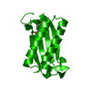
 Movie
Movie Controller


+ Open data

- Basic information
| Entry | Database: PDB / ID: 4q3f | ||||||
|---|---|---|---|---|---|---|---|
| Title | Human D-DT complexed with tartrate | ||||||
 Components Components | D-dopachrome decarboxylase | ||||||
 Keywords Keywords | LYASE / Tautomerase / cd74 | ||||||
| Function / homology |  Function and homology information Function and homology informationD-dopachrome decarboxylase / D-dopachrome decarboxylase activity / dopachrome isomerase activity / melanin biosynthetic process / phenylpyruvate tautomerase activity / cytokine receptor binding / negative regulation of macrophage chemotaxis / positive regulation of tumor necrosis factor production / positive regulation of inflammatory response / positive regulation of ERK1 and ERK2 cascade ...D-dopachrome decarboxylase / D-dopachrome decarboxylase activity / dopachrome isomerase activity / melanin biosynthetic process / phenylpyruvate tautomerase activity / cytokine receptor binding / negative regulation of macrophage chemotaxis / positive regulation of tumor necrosis factor production / positive regulation of inflammatory response / positive regulation of ERK1 and ERK2 cascade / : / extracellular exosome / cytoplasm Similarity search - Function | ||||||
| Biological species |  Homo sapiens (human) Homo sapiens (human) | ||||||
| Method |  X-RAY DIFFRACTION / MOLECULAR REPLACEMENT / Resolution: 1.8 Å X-RAY DIFFRACTION / MOLECULAR REPLACEMENT / Resolution: 1.8 Å | ||||||
 Authors Authors | Rajasekaran, D. / Lolis, E. | ||||||
 Citation Citation | Journal: Faseb J. / Year: 2014 Title: Targeting distinct tautomerase sites of D-DT and MIF with a single molecule for inhibition of neutrophil lung recruitment. Authors: Rajasekaran, D. / Zierow, S. / Syed, M. / Bucala, R. / Bhandari, V. / Lolis, E.J. | ||||||
| History |
|
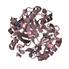
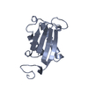
- Structure visualization
| Structure viewer | Molecule: MolmilJmol/JSmol |
|---|
- Downloads & links
-Download
| PDBx/mmCIF format | 4q3f.cif.gz | 89.8 KB | Display | PDBx/mmCIF format |
|---|---|---|---|---|
| PDB format | pdb4q3f.ent.gz | 68.8 KB | Display | PDB format |
| PDBx/mmJSON format | 4q3f.json.gz | Tree view | PDBx/mmJSON format | |
| Others |  Other downloads Other downloads |
-Validation report
| Arichive directory | https://data.pdbj.org/pub/pdb/validation_reports/q3/4q3fftp://data.pdbj.org/pub/pdb/validation_reports/q3/4q3f | HTTPS FTP |
|---|
-Related structure data


-Links
 PDBj
PDBj
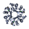
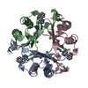
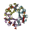
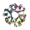
- Assembly
| Deposited unit | 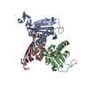
| |||||||||||||||||||||||||||||||||
|---|---|---|---|---|---|---|---|---|---|---|---|---|---|---|---|---|---|---|---|---|---|---|---|---|---|---|---|---|---|---|---|---|---|---|
| 1 | 
| |||||||||||||||||||||||||||||||||
| 2 | 
| |||||||||||||||||||||||||||||||||
| 3 | 
| |||||||||||||||||||||||||||||||||
| Unit cell |
| |||||||||||||||||||||||||||||||||
| Components on special symmetry positions |
|
-Components
| #1: Protein | Mass: 12593.526 Da / Num. of mol.: 3 Source method: isolated from a genetically manipulated source Source: (gene. exp.) Homo sapiens (human) / Gene: DDT / Production host:  #2: Chemical |   Mass: 150.087 Da / Num. of mol.: 3 / Source method: obtained synthetically / Formula: C4H6O6 Mass: 150.087 Da / Num. of mol.: 3 / Source method: obtained synthetically / Formula: C4H6O6#3: Water | ChemComp-HOH / |  Mass: 18.015 Da / Num. of mol.: 544 / Source method: isolated from a natural source / Formula: H2O Mass: 18.015 Da / Num. of mol.: 544 / Source method: isolated from a natural source / Formula: H2O |
|---|
-Experimental details
-Experiment
| Experiment | Method: X-RAY DIFFRACTION / Number of used crystals: 1 |
|---|
- Sample preparation
| Crystal | Density Matthews: 2.17 Å3/Da / Density % sol: 43.39 % |
|---|---|
| Crystal grow | Temperature: 295 K / Method: vapor diffusion, hanging drop / pH: 7.3 Details: 0.2 M Sodium tartrate dibasic dihydrate and 20% w/v Polyethylene glycol 3,350, pH 7.3, VAPOR DIFFUSION, HANGING DROP, temperature 295K |
-Data collection
| Diffraction | Mean temperature: 200 K |
|---|---|
| Diffraction source | Source: ROTATING ANODE / Type: Cu FINE FOCUS / Wavelength: 1.5418 Å |
| Detector | Type: RIGAKU RAXIS IV++ / Detector: IMAGE PLATE / Date: Mar 18, 2014 |
| Radiation | Monochromator: yale mirrors / Protocol: SINGLE WAVELENGTH / Monochromatic (M) / Laue (L): M / Scattering type: x-ray |
| Radiation wavelength | Wavelength: 1.5418 Å / Relative weight: 1 |
| Reflection | Resolution: 1.8→29.13 Å / Num. all: 74442 / Num. obs: 27672 / % possible obs: 93.8 % |
- Processing
| Software |
| |||||||||||||||||||||||||||||||||||||||||||||||||||||||||||||||||||||||||||||||||||||||||||||||||||||||||
|---|---|---|---|---|---|---|---|---|---|---|---|---|---|---|---|---|---|---|---|---|---|---|---|---|---|---|---|---|---|---|---|---|---|---|---|---|---|---|---|---|---|---|---|---|---|---|---|---|---|---|---|---|---|---|---|---|---|---|---|---|---|---|---|---|---|---|---|---|---|---|---|---|---|---|---|---|---|---|---|---|---|---|---|---|---|---|---|---|---|---|---|---|---|---|---|---|---|---|---|---|---|---|---|---|---|---|
| Refinement | Method to determine structure: MOLECULAR REPLACEMENT / Resolution: 1.8→29.13 Å / Cor.coef. Fo:Fc: 0.964 / Cor.coef. Fo:Fc free: 0.937 / SU B: 2.517 / SU ML: 0.08 / Cross valid method: THROUGHOUT / ESU R: 0.132 / ESU R Free: 0.13 / Stereochemistry target values: MAXIMUM LIKELIHOOD / Details: HYDROGENS HAVE BEEN ADDED IN THE RIDING POSITIONS
| |||||||||||||||||||||||||||||||||||||||||||||||||||||||||||||||||||||||||||||||||||||||||||||||||||||||||
| Solvent computation | Ion probe radii: 0.8 Å / Shrinkage radii: 0.8 Å / VDW probe radii: 1.2 Å / Solvent model: MASK | |||||||||||||||||||||||||||||||||||||||||||||||||||||||||||||||||||||||||||||||||||||||||||||||||||||||||
| Displacement parameters | Biso mean: 14.236 Å2
| |||||||||||||||||||||||||||||||||||||||||||||||||||||||||||||||||||||||||||||||||||||||||||||||||||||||||
| Refinement step | Cycle: LAST / Resolution: 1.8→29.13 Å
| |||||||||||||||||||||||||||||||||||||||||||||||||||||||||||||||||||||||||||||||||||||||||||||||||||||||||
| Refine LS restraints |
| |||||||||||||||||||||||||||||||||||||||||||||||||||||||||||||||||||||||||||||||||||||||||||||||||||||||||
| LS refinement shell | Resolution: 1.8→1.847 Å / Total num. of bins used: 20
|
