Temperature: 293 K / Method: vapor diffusion, sitting drop / pH: 7.5 Details: 18% PEG 10K, 0.1 M HEPES pH 7.5, MG and ADP, both at 5 mM concentrations, VAPOR DIFFUSION,SITTING DROP,NANODROP, temperature 293K, VAPOR DIFFUSION, SITTING DROP
Resolution: 2.5→44.112 Å / Num. obs: 19457 / % possible obs: 99.1 % / Observed criterion σ(I): -3 / Biso Wilson estimate: 87.218 Å2 / Rmerge(I) obs: 0.031 / Net I/σ(I): 27.12
Reflection shell
Resolution (Å)
Rmerge(I) obs
Mean I/σ(I) obs
Num. measured obs
Num. unique obs
Diffraction-ID
% possible all
2.5-2.6
0.011
2.1
14784
2102
1,2
99
2.6-2.69
0.011
3.5
12372
1660
1,2
99.5
2.69-2.8
0.011
5.7
12125
1714
1,2
98.7
2.8-2.93
0.011
8.8
12931
1738
1,2
99.5
2.93-3.08
0.011
12.5
12787
1668
1,2
99.6
3.08-3.27
0.011
18.4
12662
1706
1,2
99.8
3.27-3.52
0.011
30
12531
1722
1,2
99.4
3.52-3.88
0.011
41.6
11686
1773
1,2
99.2
3.88-4.43
0.011
54.4
12119
1712
1,2
99.7
4.43-5.57
0.011
57.3
11194
1778
1,2
98.6
5.57-44.11
0.011
63.6
12066
1884
1,2
97.7
-
Phasing
Phasing
Method: MAD
-
Processing
Software
Name
Version
Classification
NB
MolProbity
3beta29
modelbuilding
PDB_EXTRACT
3.1
dataextraction
SHELX
phasing
SHARP
phasing
XSCALE
December6, 2010
datascaling
BUSTER-TNT
2.10.0
refinement
XDS
datareduction
SHELXD
phasing
BUSTER
2.10.0
refinement
Refinement
Method to determine structure: MAD / Resolution: 2.5→44.112 Å / Cor.coef. Fo:Fc: 0.958 / Cor.coef. Fo:Fc free: 0.9501 / Occupancy max: 1 / Occupancy min: 0.5 / Cross valid method: THROUGHOUT / σ(F): 0 Details: 1. ATOM RECORD CONTAINS SUM OF TLS AND RESIDUAL B FACTORS. ANISOU R CONTAINS SUM OF TLS AND RESIDUAL U FACTORS. 2. NCS RESTRAINTS WERE APPLIED USING BUSTER'S LSSR RESTRAINT (AUTONCS). 3. DUE ...Details: 1. ATOM RECORD CONTAINS SUM OF TLS AND RESIDUAL B FACTORS. ANISOU R CONTAINS SUM OF TLS AND RESIDUAL U FACTORS. 2. NCS RESTRAINTS WERE APPLIED USING BUSTER'S LSSR RESTRAINT (AUTONCS). 3. DUE TO AMBIGUITY IN THE DENSITY OF THE LINKER 570-573, THE N-TERMINAL FRAGMENT OF EACH MONOMER CANNOT BE ASSIGNED UNIQUELY TO ONE MONOMER. THE CHAIN ASSIGNMENT WAS CHOSEN SUCH THAT THE CONNECTIVITY IN A MONOMER IS THE SAME AS OTHER HOMOLOGOUS HISTIDINE KINASES.
In the structure databanks used in Yorodumi, some data are registered as the other names, "COVID-19 virus" and "2019-nCoV". Here are the details of the virus and the list of structure data.
Jan 31, 2019. EMDB accession codes are about to change! (news from PDBe EMDB page)
EMDB accession codes are about to change! (news from PDBe EMDB page)
The allocation of 4 digits for EMDB accession codes will soon come to an end. Whilst these codes will remain in use, new EMDB accession codes will include an additional digit and will expand incrementally as the available range of codes is exhausted. The current 4-digit format prefixed with “EMD-” (i.e. EMD-XXXX) will advance to a 5-digit format (i.e. EMD-XXXXX), and so on. It is currently estimated that the 4-digit codes will be depleted around Spring 2019, at which point the 5-digit format will come into force.
The EM Navigator/Yorodumi systems omit the EMD- prefix.
Related info.:Q: What is EMD? / ID/Accession-code notation in Yorodumi/EM Navigator
Yorodumi is a browser for structure data from EMDB, PDB, SASBDB, etc.
This page is also the successor to EM Navigator detail page, and also detail information page/front-end page for Omokage search.
The word "yorodu" (or yorozu) is an old Japanese word meaning "ten thousand". "mi" (miru) is to see.
Related info.:EMDB / PDB / SASBDB / Comparison of 3 databanks / Yorodumi Search / Aug 31, 2016. New EM Navigator & Yorodumi / Yorodumi Papers / Jmol/JSmol / Function and homology information / Changes in new EM Navigator and Yorodumi
 Movie
Movie Controller

 Yorodumi
 Open data
 Basic information
 Components
 Keywords
 Function and homology information
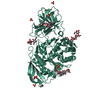
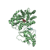
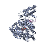
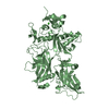
 Caulobacter crescentus (bacteria)
 X-RAY DIFFRACTION /
  Authors
 Citation
 Structure visualization
 Downloads & links
 Other downloads
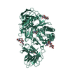


 PDBj
PDBj

 Assembly
 Mass: 18.015 Da / Num. of mol.: 19 / Source method: isolated from a natural source / Formula: H2O
 Sample preparation
 Processing