 Movie
Movie Controller

[English] 日本語
 Yorodumi
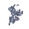
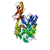
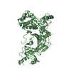
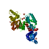
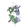
- PDB-4pz6: PCE1 guanylyltransferase bound to SER2/SER5 phosphorylated RNA po... -
+ Open data

- Basic information
| Entry | Database: PDB / ID: 4pz6 | ||||||
|---|---|---|---|---|---|---|---|
| Title | PCE1 guanylyltransferase bound to SER2/SER5 phosphorylated RNA pol II CTD | ||||||
 Components Components |
| ||||||
 Keywords Keywords | TRANSFERASE/TRANSCRIPTION / Nucleotidyl transferase / RNA capping enzyme / RNA polymerase II / Spt5 / guanylation / nuclear / TRANSFERASE-TRANSCRIPTION complex | ||||||
| Function / homology |  Function and homology information Function and homology informationRNA polymerase II, holoenzyme / RNA polymerase II transcribes snRNA genes / Processing of Capped Intron-Containing Pre-mRNA / RNA Polymerase II Promoter Escape / RNA Polymerase II Transcription Pre-Initiation And Promoter Opening / RNA Polymerase II Transcription Initiation / RNA Polymerase II Transcription Initiation And Promoter Clearance / mRNA Capping / RNA Pol II CTD phosphorylation and interaction with CE / Formation of the Early Elongation Complex ...RNA polymerase II, holoenzyme / RNA polymerase II transcribes snRNA genes / Processing of Capped Intron-Containing Pre-mRNA / RNA Polymerase II Promoter Escape / RNA Polymerase II Transcription Pre-Initiation And Promoter Opening / RNA Polymerase II Transcription Initiation / RNA Polymerase II Transcription Initiation And Promoter Clearance / mRNA Capping / RNA Pol II CTD phosphorylation and interaction with CE / Formation of the Early Elongation Complex / RNA Polymerase II Pre-transcription Events / TP53 Regulates Transcription of DNA Repair Genes / Estrogen-dependent gene expression / mRNA Splicing - Major Pathway / Transcriptional regulation by small RNAs / Formation of TC-NER Pre-Incision Complex / Dual incision in TC-NER / Gap-filling DNA repair synthesis and ligation in TC-NER / co-transcriptional lncRNA 3' end processing, cleavage and polyadenylation pathway / intracellular phosphate ion homeostasis / chromatin-protein adaptor activity / termination of RNA polymerase II transcription / RNA polymerase II C-terminal domain binding / 7-methylguanosine mRNA capping / RNA polymerase II, core complex / transcription initiation at RNA polymerase II promoter / transcription elongation by RNA polymerase II / mRNA transcription by RNA polymerase II / DNA-directed RNA polymerase / DNA-directed RNA polymerase activity / mRNA guanylyltransferase / mRNA guanylyltransferase activity / chromatin / GTP binding / DNA binding / ATP binding / metal ion binding / nucleus Similarity search - Function | ||||||
| Biological species |  | ||||||
| Method |  X-RAY DIFFRACTION / SYNCHROTRON / MOLECULAR REPLACEMENT / Resolution: 2.406 Å X-RAY DIFFRACTION / SYNCHROTRON / MOLECULAR REPLACEMENT / Resolution: 2.406 Å | ||||||
 Authors Authors | Doamekpor, S.K. / Lima, C.D. | ||||||
 Citation Citation | Journal: Genes Dev. / Year: 2014 Title: How an mRNA capping enzyme reads distinct RNA polymerase II and Spt5 CTD phosphorylation codes. Authors: Doamekpor, S.K. / Sanchez, A.M. / Schwer, B. / Shuman, S. / Lima, C.D. | ||||||
| History |
|
- Structure visualization
| Structure viewer | Molecule: MolmilJmol/JSmol |
|---|
- Downloads & links
-Download
| PDBx/mmCIF format | 4pz6.cif.gz | 174.6 KB | Display | PDBx/mmCIF format |
|---|---|---|---|---|
| PDB format | pdb4pz6.ent.gz | 137.1 KB | Display | PDB format |
| PDBx/mmJSON format | 4pz6.json.gz | Tree view | PDBx/mmJSON format | |
| Others |  Other downloads Other downloads |
-Validation report
| Arichive directory | https://data.pdbj.org/pub/pdb/validation_reports/pz/4pz6ftp://data.pdbj.org/pub/pdb/validation_reports/pz/4pz6 | HTTPS FTP |
|---|
-Related structure data
| Related structure data |  4pz7C 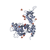 4pz8C 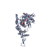 1p16S C: citing same article ( S: Starting model for refinement |
|---|---|
| Similar structure data |


-Links
 PDBj
PDBj


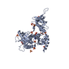
- Assembly
| Deposited unit | 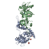
| ||||||||
|---|---|---|---|---|---|---|---|---|---|
| 1 | 
| ||||||||
| 2 | 
| ||||||||
| Unit cell |
|
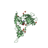
-Components
| #1: Protein | Mass: 47034.062 Da / Num. of mol.: 2 Source method: isolated from a genetically manipulated source Source: (gene. exp.) Strain: 972 / ATCC 24843 / Gene: ceg1, ceg1/pce1, pce1, SPBC2F12.08c / Plasmid: pSMT3 (SUMO) / Production host:  #2: Protein/peptide | Mass: 2657.111 Da / Num. of mol.: 2 Fragment: phosphorylated C-terminal domain peptide (UNP residues 1578-1598) Source method: obtained synthetically / Source: (synth.) #3: Chemical | ChemComp-GMP / | 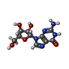  Mass: 283.241 Da / Num. of mol.: 1 / Source method: obtained synthetically / Formula: C10H13N5O5 Mass: 283.241 Da / Num. of mol.: 1 / Source method: obtained synthetically / Formula: C10H13N5O5#4: Water | ChemComp-HOH / |  Mass: 18.015 Da / Num. of mol.: 212 / Source method: isolated from a natural source / Formula: H2O Mass: 18.015 Da / Num. of mol.: 212 / Source method: isolated from a natural source / Formula: H2OHas protein modification | Y | |
|---|
-Experimental details
-Experiment
| Experiment | Method: X-RAY DIFFRACTION / Number of used crystals: 1 |
|---|
- Sample preparation
| Crystal | Density Matthews: 2.58 Å3/Da / Density % sol: 52.34 % |
|---|---|
| Crystal grow | Temperature: 291 K / Method: vapor diffusion, hanging drop / pH: 7 Details: 23% PEG400, 0.1 M sodium acetate, pH 7.0, VAPOR DIFFUSION, HANGING DROP, temperature 291K |
-Data collection
| Diffraction | Mean temperature: 100 K |
|---|---|
| Diffraction source | Source: SYNCHROTRON / Site: APS  / Beamline: 24-ID-E / Wavelength: 0.979 Å / Beamline: 24-ID-E / Wavelength: 0.979 Å |
| Detector | Type: ADSC QUANTUM 315 / Detector: CCD / Date: Aug 5, 2009 |
| Radiation | Monochromator: Si(220) side bounce / Protocol: SINGLE WAVELENGTH / Monochromatic (M) / Laue (L): M / Scattering type: x-ray |
| Radiation wavelength | Wavelength: 0.979 Å / Relative weight: 1 |
| Reflection | Resolution: 2.406→50 Å / Num. all: 39406 / Num. obs: 38342 / % possible obs: 97.3 % / Observed criterion σ(F): 0 / Observed criterion σ(I): 0 / Redundancy: 3.9 % / Biso Wilson estimate: 45.5 Å2 / Rmerge(I) obs: 0.092 / Net I/σ(I): 9.6 |
| Reflection shell | Resolution: 2.406→2.49 Å / Redundancy: 2.3 % / Rmerge(I) obs: 0.41 / Mean I/σ(I) obs: 2.9 / Num. unique all: 3511 / % possible all: 89.4 |
- Processing
| Software |
| |||||||||||||||||||||||||||||||||||||||||||||||||||||||||||||||||||||||||||||||||||||||||||||||||||||||||
|---|---|---|---|---|---|---|---|---|---|---|---|---|---|---|---|---|---|---|---|---|---|---|---|---|---|---|---|---|---|---|---|---|---|---|---|---|---|---|---|---|---|---|---|---|---|---|---|---|---|---|---|---|---|---|---|---|---|---|---|---|---|---|---|---|---|---|---|---|---|---|---|---|---|---|---|---|---|---|---|---|---|---|---|---|---|---|---|---|---|---|---|---|---|---|---|---|---|---|---|---|---|---|---|---|---|---|
| Refinement | Method to determine structure: MOLECULAR REPLACEMENT Starting model: PDB ENTRY 1P16 Resolution: 2.406→36.274 Å / SU ML: 0.29 / Cross valid method: THROUGHOUT / σ(F): 1.43 / Phase error: 24.78 / Stereochemistry target values: Engh & Huber
| |||||||||||||||||||||||||||||||||||||||||||||||||||||||||||||||||||||||||||||||||||||||||||||||||||||||||
| Solvent computation | Shrinkage radii: 0.9 Å / VDW probe radii: 1.11 Å / Solvent model: FLAT BULK SOLVENT MODEL | |||||||||||||||||||||||||||||||||||||||||||||||||||||||||||||||||||||||||||||||||||||||||||||||||||||||||
| Refinement step | Cycle: LAST / Resolution: 2.406→36.274 Å
| |||||||||||||||||||||||||||||||||||||||||||||||||||||||||||||||||||||||||||||||||||||||||||||||||||||||||
| Refine LS restraints |
| |||||||||||||||||||||||||||||||||||||||||||||||||||||||||||||||||||||||||||||||||||||||||||||||||||||||||
| LS refinement shell |
|
