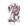
| Entry | Database: PDB / ID: 4pyw
|
|---|
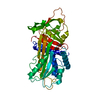
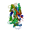
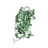
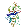
| Title | 1.92 angstrom crystal structure of A1AT:TTAI ternary complex |
|---|
 Components Components | - ACE-THR-THR-ALA-ILE-NH2
- Alpha-1-antitrypsin
|
|---|
 Keywords Keywords | Hydrolase inhibitor / Serpin |
|---|
| Function / homology |  Function and homology information Function and homology information
Cargo concentration in the ER / COPII-coated ER to Golgi transport vesicle / COPII-mediated vesicle transport / endoplasmic reticulum-Golgi intermediate compartment membrane / platelet alpha granule lumen / acute-phase response / Post-translational protein phosphorylation / serine-type endopeptidase inhibitor activity / Regulation of Insulin-like Growth Factor (IGF) transport and uptake by Insulin-like Growth Factor Binding Proteins (IGFBPs) / blood coagulation ...Cargo concentration in the ER / COPII-coated ER to Golgi transport vesicle / COPII-mediated vesicle transport / endoplasmic reticulum-Golgi intermediate compartment membrane / platelet alpha granule lumen / acute-phase response / Post-translational protein phosphorylation / serine-type endopeptidase inhibitor activity / Regulation of Insulin-like Growth Factor (IGF) transport and uptake by Insulin-like Growth Factor Binding Proteins (IGFBPs) / blood coagulation / Platelet degranulation / extracellular matrix / protease binding / ficolin-1-rich granule lumen / endoplasmic reticulum lumen / Neutrophil degranulation / Golgi apparatus / endoplasmic reticulum / : / extracellular exosome / extracellular region / identical protein bindingSimilarity search - Function Antithrombin; Chain I, domain 2 / Antithrombin, subunit I, domain 2 / Alpha-1-antitrypsin; domain 1 / Alpha-1-antitrypsin, domain 1 / Serpin, conserved site / Serpins signature. / Serpin superfamily, domain 2 / Serpin family / Serpin domain / Serpin superfamily ...Antithrombin; Chain I, domain 2 / Antithrombin, subunit I, domain 2 / Alpha-1-antitrypsin; domain 1 / Alpha-1-antitrypsin, domain 1 / Serpin, conserved site / Serpins signature. / Serpin superfamily, domain 2 / Serpin family / Serpin domain / Serpin superfamily / Serpin superfamily, domain 1 / Serpin (serine protease inhibitor) / SERine Proteinase INhibitors / Roll / 2-Layer Sandwich / Mainly Beta / Alpha BetaSimilarity search - Domain/homology |
|---|
| Biological species |  Homo sapiens (human) Homo sapiens (human)
synthetic construct (others) |
|---|
| Method |  X-RAY DIFFRACTION / SYNCHROTRON / MOLECULAR REPLACEMENT / Resolution: 1.91 Å X-RAY DIFFRACTION / SYNCHROTRON / MOLECULAR REPLACEMENT / Resolution: 1.91 Å |
|---|
 Authors Authors | Nyon, M.P. / Day, J. / Gooptu, B. |
|---|
 Citation Citation | Journal: Protein Sci. / Year: 2015
Title: An integrative approach combining ion mobility mass spectrometry, X-ray crystallography, and nuclear magnetic resonance spectroscopy to study the conformational dynamics of alpha 1 - ...Title: An integrative approach combining ion mobility mass spectrometry, X-ray crystallography, and nuclear magnetic resonance spectroscopy to study the conformational dynamics of alpha 1 -antitrypsin upon ligand binding.
Authors: Nyon, M.P. / Prentice, T. / Day, J. / Kirkpatrick, J. / Sivalingam, G.N. / Levy, G. / Haq, I. / Irving, J.A. / Lomas, D.A. / Christodoulou, J. / Gooptu, B. / Thalassinos, K. |
|---|
| History | | Deposition | Mar 28, 2014 | Deposition site: RCSB / Processing site: RCSB |
|---|
| Revision 1.0 | Jun 10, 2015 | Provider: repository / Type: Initial release |
|---|
| Revision 1.1 | Aug 24, 2022 | Group: Database references / Derived calculations
Category: citation / database_2 ...citation / database_2 / struct_conn / struct_ref_seq_dif / struct_site
Item: _citation.journal_volume / _citation.page_first ..._citation.journal_volume / _citation.page_first / _citation.page_last / _citation.title / _database_2.pdbx_DOI / _database_2.pdbx_database_accession / _struct_conn.pdbx_leaving_atom_flag / _struct_ref_seq_dif.details / _struct_site.pdbx_auth_asym_id / _struct_site.pdbx_auth_comp_id / _struct_site.pdbx_auth_seq_id |
|---|
| Revision 1.2 | Oct 30, 2024 | Group: Data collection / Structure summary
Category: chem_comp_atom / chem_comp_bond ...chem_comp_atom / chem_comp_bond / pdbx_entry_details / pdbx_modification_feature |
|---|
|
|---|
 Movie
Movie Controller


 Open data
 Basic information
 Structure visualization
 Downloads & links
 Other downloads


 PDBj
PDBj

 Assembly


 Mass: 92.094 Da / Num. of mol.: 2 / Source method: obtained synthetically / Formula: C3H8O3
 Mass: 18.015 Da / Num. of mol.: 96 / Source method: isolated from a natural source / Formula: H2O
 Sample preparation
 / Beamline: ID23-2 / Wavelength: 1.0332 Å
 Processing