 Movie
Movie Controller

[English] 日本語
 Yorodumi
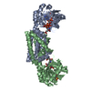
- PDB-4p6q: The crystal structure of the Split End protein SHARP adds a new l... -
+ Open data

- Basic information
| Entry | Database: PDB / ID: 4p6q | |||||||||
|---|---|---|---|---|---|---|---|---|---|---|
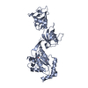
| Title | The crystal structure of the Split End protein SHARP adds a new layer of complexity to proteins containing RNA Recognition Motifs | |||||||||
 Components Components | Msx2-interacting protein | |||||||||
 Keywords Keywords | TRANSCRIPTION / RNA-recognition Motif / SPEN protein / Steroid RNA Activator / Transcriptional Regulation | |||||||||
| Function / homology |  Function and homology information Function and homology informationrandom inactivation of X chromosome / regulatory ncRNA-mediated heterochromatin formation / RHOBTB1 GTPase cycle / positive regulation of neurogenesis / Notch signaling pathway / transcription repressor complex / transcription corepressor activity / RNA polymerase II-specific DNA-binding transcription factor binding / negative regulation of DNA-templated transcription / mRNA binding ...random inactivation of X chromosome / regulatory ncRNA-mediated heterochromatin formation / RHOBTB1 GTPase cycle / positive regulation of neurogenesis / Notch signaling pathway / transcription repressor complex / transcription corepressor activity / RNA polymerase II-specific DNA-binding transcription factor binding / negative regulation of DNA-templated transcription / mRNA binding / regulation of transcription by RNA polymerase II / negative regulation of transcription by RNA polymerase II / DNA binding / RNA binding / extracellular exosome / nucleoplasm / nucleus Similarity search - Function | |||||||||
| Biological species |  Homo sapiens (human) Homo sapiens (human) | |||||||||
| Method |  X-RAY DIFFRACTION / SYNCHROTRON / MOLECULAR REPLACEMENT / Resolution: 2 Å X-RAY DIFFRACTION / SYNCHROTRON / MOLECULAR REPLACEMENT / Resolution: 2 Å | |||||||||
 Authors Authors | Arieti, F. / Gabus, C. / Tambalo, M. / Huet, T. / Round, A. / Thore, S. | |||||||||
| Funding support |  Switzerland, 1items Switzerland, 1items
| |||||||||
 Citation Citation | Journal: Nucleic Acids Res. / Year: 2014 Title: The crystal structure of the Split End protein SHARP adds a new layer of complexity to proteins containing RNA recognition motifs. Authors: Arieti, F. / Gabus, C. / Tambalo, M. / Huet, T. / Round, A. / Thore, S. | |||||||||
| History |
|
- Structure visualization
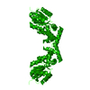
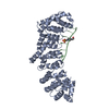
| Structure viewer | Molecule: MolmilJmol/JSmol |
|---|
- Downloads & links
-Download
| PDBx/mmCIF format | 4p6q.cif.gz | 137.5 KB | Display | PDBx/mmCIF format |
|---|---|---|---|---|
| PDB format | pdb4p6q.ent.gz | 106 KB | Display | PDB format |
| PDBx/mmJSON format | 4p6q.json.gz | Tree view | PDBx/mmJSON format | |
| Others |  Other downloads Other downloads |
-Validation report
| Arichive directory | https://data.pdbj.org/pub/pdb/validation_reports/p6/4p6qftp://data.pdbj.org/pub/pdb/validation_reports/p6/4p6q | HTTPS FTP |
|---|
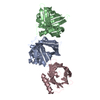
-Related structure data
| Related structure data |  1whyS  1x55S 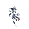 2adcS 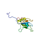 2cpeS 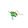 2cpzS  2cqbS  2dguS  2dnmS  2i38S 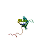 2lcwS 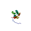 2ytcS  3md1S 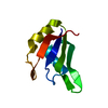 4f26S S: Starting model for refinement |
|---|---|
| Similar structure data |


-Links
 PDBj
PDBj

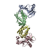
- Assembly
| Deposited unit | 
| ||||||||
|---|---|---|---|---|---|---|---|---|---|
| 1 |
| ||||||||
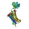
| Unit cell |
|
-Components
| #1: Protein | Mass: 33056.730 Da / Num. of mol.: 1 Source method: isolated from a genetically manipulated source Source: (gene. exp.) Homo sapiens (human) / Gene: SPEN, KIAA0929, MINT, SHARP / Plasmid: pET42a / Production host:  |
|---|---|
| #2: Chemical | ChemComp-SO4 /   Mass: 96.063 Da / Num. of mol.: 1 / Source method: obtained synthetically / Formula: SO4 Mass: 96.063 Da / Num. of mol.: 1 / Source method: obtained synthetically / Formula: SO4 |
| #3: Water | ChemComp-HOH /  Mass: 18.015 Da / Num. of mol.: 156 / Source method: isolated from a natural source / Formula: H2O Mass: 18.015 Da / Num. of mol.: 156 / Source method: isolated from a natural source / Formula: H2O |
-Experimental details
-Experiment
| Experiment | Method: X-RAY DIFFRACTION |
|---|
- Sample preparation
| Crystal | Density Matthews: 2.84 Å3/Da / Density % sol: 56.75 % |
|---|---|
| Crystal grow | Temperature: 277 K / Method: vapor diffusion, sitting drop / pH: 5.6 Details: 15% PEG 8000, 0.1M ammonium sulphate, 0.01M magnesium chloride, 0.05M 2-(N-morpholino)ethanesulfonic acid PH range: 5.4-6 |
-Data collection
| Diffraction | Mean temperature: 100 K |
|---|---|
| Diffraction source | Source: SYNCHROTRON / Site: ESRF  / Beamline: BM16 / Wavelength: 1 Å / Beamline: BM16 / Wavelength: 1 Å |
| Detector | Type: ADSC QUANTUM 315r / Detector: CCD / Date: Nov 29, 2012 |
| Radiation | Protocol: SINGLE WAVELENGTH / Monochromatic (M) / Laue (L): M / Scattering type: x-ray |
| Radiation wavelength | Wavelength: 1 Å / Relative weight: 1 |
| Reflection | Resolution: 2→28.71 Å / Num. obs: 26164 / % possible obs: 99.67 % / Redundancy: 5.38 % / Net I/σ(I): 20.44 |
| Reflection shell | Highest resolution: 2 Å / Redundancy: 5.63 % / % possible all: 100 |
- Processing
| Software |
| ||||||||||||||||||||||||||||||||||||||||||||||||||||||||||||||||||||||||||||||||||||||||||||||||||||||||||||||||||||||||||||||||||||||||||||||||||||||||||||
|---|---|---|---|---|---|---|---|---|---|---|---|---|---|---|---|---|---|---|---|---|---|---|---|---|---|---|---|---|---|---|---|---|---|---|---|---|---|---|---|---|---|---|---|---|---|---|---|---|---|---|---|---|---|---|---|---|---|---|---|---|---|---|---|---|---|---|---|---|---|---|---|---|---|---|---|---|---|---|---|---|---|---|---|---|---|---|---|---|---|---|---|---|---|---|---|---|---|---|---|---|---|---|---|---|---|---|---|---|---|---|---|---|---|---|---|---|---|---|---|---|---|---|---|---|---|---|---|---|---|---|---|---|---|---|---|---|---|---|---|---|---|---|---|---|---|---|---|---|---|---|---|---|---|---|---|---|---|
| Refinement | Method to determine structure: MOLECULAR REPLACEMENT Starting model: Ensemble 1 (PDB codes: 3MD1, 2ADC, 2DNM, 2DGU, 2CQB); Ensemble 2 (PDB codes: X4AR, 2YTC, 4F26, 1WHY, 2CPZ); Ensemble 3 (PDB codes: 2I38, 1WHY, 1X55, 2LCW, 2CPE). ...Starting model: Ensemble 1 (PDB codes: 3MD1, 2ADC, 2DNM, 2DGU, 2CQB); Ensemble 2 (PDB codes: X4AR, 2YTC, 4F26, 1WHY, 2CPZ); Ensemble 3 (PDB codes: 2I38, 1WHY, 1X55, 2LCW, 2CPE). Resolution: 2→28.71 Å / SU ML: 0.26 / Cross valid method: THROUGHOUT / σ(F): 2 / Phase error: 25.01 / Stereochemistry target values: ML
| ||||||||||||||||||||||||||||||||||||||||||||||||||||||||||||||||||||||||||||||||||||||||||||||||||||||||||||||||||||||||||||||||||||||||||||||||||||||||||||
| Solvent computation | Shrinkage radii: 0.9 Å / VDW probe radii: 1.11 Å / Solvent model: FLAT BULK SOLVENT MODEL | ||||||||||||||||||||||||||||||||||||||||||||||||||||||||||||||||||||||||||||||||||||||||||||||||||||||||||||||||||||||||||||||||||||||||||||||||||||||||||||
| Refinement step | Cycle: LAST / Resolution: 2→28.71 Å
| ||||||||||||||||||||||||||||||||||||||||||||||||||||||||||||||||||||||||||||||||||||||||||||||||||||||||||||||||||||||||||||||||||||||||||||||||||||||||||||
| Refine LS restraints |
| ||||||||||||||||||||||||||||||||||||||||||||||||||||||||||||||||||||||||||||||||||||||||||||||||||||||||||||||||||||||||||||||||||||||||||||||||||||||||||||
| LS refinement shell |
| ||||||||||||||||||||||||||||||||||||||||||||||||||||||||||||||||||||||||||||||||||||||||||||||||||||||||||||||||||||||||||||||||||||||||||||||||||||||||||||
| Refinement TLS params. | Method: refined / Refine-ID: X-RAY DIFFRACTION
| ||||||||||||||||||||||||||||||||||||||||||||||||||||||||||||||||||||||||||||||||||||||||||||||||||||||||||||||||||||||||||||||||||||||||||||||||||||||||||||
| Refinement TLS group |
|
