 Movie
Movie Controller

[English] 日本語
 Yorodumi
- PDB-4p5a: Crystal structure of a UMP/dUMP methylase PolB from Streptomyces ... -
+ Open data

- Basic information
| Entry | Database: PDB / ID: 4p5a | ||||||
|---|---|---|---|---|---|---|---|
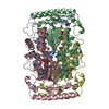
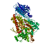
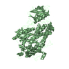
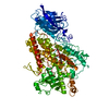
| Title | Crystal structure of a UMP/dUMP methylase PolB from Streptomyces cacaoi bound with 5-Br UMP | ||||||
 Components Components | Thymidylate synthase ThyX | ||||||
 Keywords Keywords | TRANSFERASE / Tetramer / UMP/dUMP methylase / ThyX homolog | ||||||
| Function / homology |  Function and homology information Function and homology informationthymidylate synthase (FAD) / thymidylate synthase (FAD) activity / thymidylate synthase activity / dTTP biosynthetic process / dTMP biosynthetic process / NADPH binding / flavin adenine dinucleotide binding / methylation Similarity search - Function | ||||||
| Biological species |  Streptomyces cacaoi subsp. asoensis (bacteria) Streptomyces cacaoi subsp. asoensis (bacteria) | ||||||
| Method |  X-RAY DIFFRACTION / SYNCHROTRON / Resolution: 1.76 Å X-RAY DIFFRACTION / SYNCHROTRON / Resolution: 1.76 Å | ||||||
 Authors Authors | Li, Y. / Chen, W. / Li, J. / Xia, Z. / Deng, Z. / Zhou, J. | ||||||
 Citation Citation | Journal: To Be Published Title: Crystal structure of a UMP/dUMP methylase PolB from Streptomyces cacaoi with 5-Br UMP Authors: Li, Y. / Chen, W. / Li, J. / Xia, Z. / Deng, Z. / Zhou, J. | ||||||
| History |
|
- Structure visualization
| Structure viewer | Molecule: MolmilJmol/JSmol |
|---|
- Downloads & links
-Download
| PDBx/mmCIF format | 4p5a.cif.gz | 213.7 KB | Display | PDBx/mmCIF format |
|---|---|---|---|---|
| PDB format | pdb4p5a.ent.gz | 171.5 KB | Display | PDB format |
| PDBx/mmJSON format | 4p5a.json.gz | Tree view | PDBx/mmJSON format | |
| Others |  Other downloads Other downloads |
-Validation report
| Arichive directory | https://data.pdbj.org/pub/pdb/validation_reports/p5/4p5aftp://data.pdbj.org/pub/pdb/validation_reports/p5/4p5a | HTTPS FTP |
|---|
-Related structure data
| Related structure data |  4p5c |
|---|---|
| Similar structure data |


-Links
 PDBj
PDBj- Assembly
| Deposited unit | 
| ||||||||
|---|---|---|---|---|---|---|---|---|---|
| 1 |
| ||||||||
| Unit cell |
|
-Components
| #1: Protein | Mass: 27126.588 Da / Num. of mol.: 4 / Fragment: UNP residues 19-257 Source method: isolated from a genetically manipulated source Source: (gene. exp.) Streptomyces cacaoi subsp. asoensis (bacteria)Gene: polB, thyX / Plasmid: pET28a / Production host: #2: Chemical | ChemComp-FAD /   Mass: 785.550 Da / Num. of mol.: 4 / Source method: obtained synthetically / Formula: C27H33N9O15P2 / Comment: FAD*YM Mass: 785.550 Da / Num. of mol.: 4 / Source method: obtained synthetically / Formula: C27H33N9O15P2 / Comment: FAD*YM#3: Chemical | ChemComp-5BU /   Type: RNA linking / Mass: 403.077 Da / Num. of mol.: 4 / Source method: obtained synthetically / Formula: C9H12BrN2O9P Type: RNA linking / Mass: 403.077 Da / Num. of mol.: 4 / Source method: obtained synthetically / Formula: C9H12BrN2O9P#4: Water | ChemComp-HOH / |  Mass: 18.015 Da / Num. of mol.: 733 / Source method: isolated from a natural source / Formula: H2O Mass: 18.015 Da / Num. of mol.: 733 / Source method: isolated from a natural source / Formula: H2O |
|---|
-Experimental details
-Experiment
| Experiment | Method: X-RAY DIFFRACTION |
|---|
- Sample preparation
| Crystal | Density Matthews: 2.48 Å3/Da / Density % sol: 50.46 % |
|---|---|
| Crystal grow | Temperature: 293 K / Method: vapor diffusion, sitting drop / Details: 0.1M HEPES, pH 7.5, 10%PEG4000, 5 % isopropanol |
-Data collection
| Diffraction | Mean temperature: 100 K |
|---|---|
| Diffraction source | Source: SYNCHROTRON / Site: SSRF  / Beamline: BL17U / Wavelength: 0.9798 Å / Beamline: BL17U / Wavelength: 0.9798 Å |
| Detector | Type: MARMOSAIC 225 mm CCD / Detector: CCD / Date: Jul 9, 2009 |
| Radiation | Protocol: SINGLE WAVELENGTH / Monochromatic (M) / Laue (L): M / Scattering type: x-ray |
| Radiation wavelength | Wavelength: 0.9798 Å / Relative weight: 1 |
| Reflection | Resolution: 1.76→50 Å / Num. obs: 103864 / % possible obs: 98.7 % / Redundancy: 7.5 % / Net I/σ(I): 13.49 |
- Processing
| Software | Name: REFMAC / Version: 5.5.0102 / Classification: refinement | ||||||||||||||||||||||||||||||||||||||||||||||||||||||||||||||||||||||||||||||||||||||||||||||||||||||||||||||||||||||||||||||||||||||||||||||||||||||||||||||||||||||||||||||||||||||
|---|---|---|---|---|---|---|---|---|---|---|---|---|---|---|---|---|---|---|---|---|---|---|---|---|---|---|---|---|---|---|---|---|---|---|---|---|---|---|---|---|---|---|---|---|---|---|---|---|---|---|---|---|---|---|---|---|---|---|---|---|---|---|---|---|---|---|---|---|---|---|---|---|---|---|---|---|---|---|---|---|---|---|---|---|---|---|---|---|---|---|---|---|---|---|---|---|---|---|---|---|---|---|---|---|---|---|---|---|---|---|---|---|---|---|---|---|---|---|---|---|---|---|---|---|---|---|---|---|---|---|---|---|---|---|---|---|---|---|---|---|---|---|---|---|---|---|---|---|---|---|---|---|---|---|---|---|---|---|---|---|---|---|---|---|---|---|---|---|---|---|---|---|---|---|---|---|---|---|---|---|---|---|---|
| Refinement | Resolution: 1.76→50 Å / Cor.coef. Fo:Fc: 0.965 / Cor.coef. Fo:Fc free: 0.949 / SU B: 1.784 / SU ML: 0.058 / Cross valid method: THROUGHOUT / ESU R: 0.098 / ESU R Free: 0.1 / Stereochemistry target values: MAXIMUM LIKELIHOOD
| ||||||||||||||||||||||||||||||||||||||||||||||||||||||||||||||||||||||||||||||||||||||||||||||||||||||||||||||||||||||||||||||||||||||||||||||||||||||||||||||||||||||||||||||||||||||
| Solvent computation | Ion probe radii: 0.8 Å / Shrinkage radii: 0.8 Å / VDW probe radii: 1.4 Å / Solvent model: MASK | ||||||||||||||||||||||||||||||||||||||||||||||||||||||||||||||||||||||||||||||||||||||||||||||||||||||||||||||||||||||||||||||||||||||||||||||||||||||||||||||||||||||||||||||||||||||
| Displacement parameters | Biso mean: 19.28 Å2
| ||||||||||||||||||||||||||||||||||||||||||||||||||||||||||||||||||||||||||||||||||||||||||||||||||||||||||||||||||||||||||||||||||||||||||||||||||||||||||||||||||||||||||||||||||||||
| Refinement step | Cycle: 1 / Resolution: 1.76→50 Å
| ||||||||||||||||||||||||||||||||||||||||||||||||||||||||||||||||||||||||||||||||||||||||||||||||||||||||||||||||||||||||||||||||||||||||||||||||||||||||||||||||||||||||||||||||||||||
| Refine LS restraints |
|