 Movie
Movie Controller


+ Open data

- Basic information
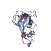
| Entry | Database: PDB / ID: 4oxf | ||||||
|---|---|---|---|---|---|---|---|
| Title | Structure of ECP in complex with citrate ions at 1.50 Angstroms | ||||||
 Components Components | Eosinophil cationic protein | ||||||
 Keywords Keywords | HYDROLASE / RNase 3 / Eosinophil Cationic Protein (ECP) | ||||||
| Function / homology |  Function and homology information Function and homology informationinduction of bacterial agglutination / Hydrolases; Acting on ester bonds; Endoribonucleases producing 3'-phosphomonoesters / RNA catabolic process / Antimicrobial peptides / RNA nuclease activity / innate immune response in mucosa / lipopolysaccharide binding / chemotaxis / azurophil granule lumen / antimicrobial humoral immune response mediated by antimicrobial peptide ...induction of bacterial agglutination / Hydrolases; Acting on ester bonds; Endoribonucleases producing 3'-phosphomonoesters / RNA catabolic process / Antimicrobial peptides / RNA nuclease activity / innate immune response in mucosa / lipopolysaccharide binding / chemotaxis / azurophil granule lumen / antimicrobial humoral immune response mediated by antimicrobial peptide / antibacterial humoral response / endonuclease activity / defense response to Gram-negative bacterium / nucleic acid binding / defense response to Gram-positive bacterium / innate immune response / hydrolase activity / Neutrophil degranulation / : / extracellular region Similarity search - Function | ||||||
| Biological species |  Homo sapiens (human) Homo sapiens (human) | ||||||
| Method |  X-RAY DIFFRACTION / SYNCHROTRON / Resolution: 1.5 Å X-RAY DIFFRACTION / SYNCHROTRON / Resolution: 1.5 Å | ||||||
 Authors Authors | Blanco, J.A. / Boix, E. / Moussaoui, M. / Salazar, V.A. | ||||||
| Funding support |  Spain, 1items Spain, 1items
| ||||||
 Citation Citation | Journal: To be published Title: Structure of ECP in complex with citrate ions at 1.50 Angstroms Authors: Blanco, J.A. / Boix, E. / Moussaoui, M. / Salazar, V.A. #1: Journal: J.Mol.Biol. / Year: 2000Title: Three-dimensional crystal structure of human eosinophil cationic protein (RNase 3) at 1.75 A resolution. Authors: Mallorqui-Fernandez, G. / Pous, J. / Peracaula, R. / Aymami, J. / Maeda, T. / Tada, H. / Yamada, H. / Seno, M. / de Llorens, R. / Gomis-Ruth, F.X. / Coll, M. #2: Journal: J.Struct.Biol. / Year: 2012Title: The sulfate-binding site structure of the human eosinophil cationic protein as revealed by a new crystal form. Authors: Boix, E. / Pulido, D. / Moussaoui, M. / Nogues, M.V. / Russi, S. | ||||||
| History |
|
- Structure visualization
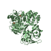
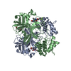
| Structure viewer | Molecule: MolmilJmol/JSmol |
|---|
- Downloads & links
-Download
| PDBx/mmCIF format | 4oxf.cif.gz | 84.3 KB | Display | PDBx/mmCIF format |
|---|---|---|---|---|
| PDB format | pdb4oxf.ent.gz | 63.3 KB | Display | PDB format |
| PDBx/mmJSON format | 4oxf.json.gz | Tree view | PDBx/mmJSON format | |
| Others |  Other downloads Other downloads |
-Validation report
| Arichive directory | https://data.pdbj.org/pub/pdb/validation_reports/ox/4oxfftp://data.pdbj.org/pub/pdb/validation_reports/ox/4oxf | HTTPS FTP |
|---|
-Related structure data
| Related structure data | |
|---|---|
| Similar structure data |


-Links
 PDBj
PDBj


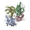
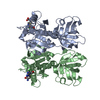
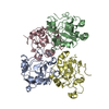
- Assembly
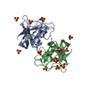
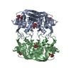
| Deposited unit | 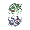
| ||||||||
|---|---|---|---|---|---|---|---|---|---|
| 1 | 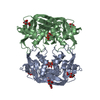
| ||||||||
| Unit cell |
|
-Components
| #1: Protein | Mass: 15730.072 Da / Num. of mol.: 2 Source method: isolated from a genetically manipulated source Source: (gene. exp.) Homo sapiens (human) / Tissue: Bone Marrow / Cell: Eosinophil / Gene: ECP,RNASE3,RNS3 / Plasmid: pET11c / Cell (production host): Bacteria / Production host:  References: UniProt: P12724, Hydrolases; Acting on ester bonds; Endoribonucleases producing 3'-phosphomonoesters #2: Chemical | ChemComp-CIT /   Mass: 192.124 Da / Num. of mol.: 5 / Source method: obtained synthetically / Formula: C6H8O7 Mass: 192.124 Da / Num. of mol.: 5 / Source method: obtained synthetically / Formula: C6H8O7#3: Chemical |   Mass: 55.845 Da / Num. of mol.: 2 / Source method: obtained synthetically / Formula: Fe Mass: 55.845 Da / Num. of mol.: 2 / Source method: obtained synthetically / Formula: Fe#4: Water | ChemComp-HOH / |  Mass: 18.015 Da / Num. of mol.: 400 / Source method: isolated from a natural source / Formula: H2O Mass: 18.015 Da / Num. of mol.: 400 / Source method: isolated from a natural source / Formula: H2OHas protein modification | Y | Nonpolymer details | AUTHORS STATE THAT BOTH IRON AND CITRATE ATOMS HAVE BEEN ADJUSTED TO THE CORRESPONDING ELECTRON ...AUTHORS STATE THAT BOTH IRON AND CITRATE ATOMS HAVE BEEN ADJUSTED TO THE CORRESPOND | |
|---|
-Experimental details
-Experiment
| Experiment | Method: X-RAY DIFFRACTION / Number of used crystals: 1 |
|---|
- Sample preparation
| Crystal | Density Matthews: 2.73 Å3/Da / Density % sol: 54.97 % |
|---|---|
| Crystal grow | Temperature: 293 K / Method: vapor diffusion, hanging drop / pH: 5.2 Details: Protein resuspended in sodium cacodylate buffer (20mM, pH5.0). 1 uL of protein sample was mixed with 1 uL of crystallisation buffer (0.1M sodium citrate pH5.2, 8% Jeffamine M-600, 10mM iron(III) chloride) PH range: 5.0 - 5.2 |
-Data collection
| Diffraction | Mean temperature: 100 K |
|---|---|
| Diffraction source | Source: SYNCHROTRON / Site: ALBA / Beamline: XALOC / Wavelength: 0.9795 Å |
| Detector | Type: DECTRIS PILATUS 6M / Detector: PIXEL / Date: Jul 10, 2013 |
| Radiation | Protocol: SINGLE WAVELENGTH / Monochromatic (M) / Laue (L): M / Scattering type: x-ray |
| Radiation wavelength | Wavelength: 0.9795 Å / Relative weight: 1 |
| Reflection | Resolution: 1.5→52.53 Å / Num. obs: 61922 / % possible obs: 99.3 % / Redundancy: 8.6 % / Rmerge(I) obs: 0.03 / Net I/σ(I): 32.3 |
| Reflection shell | Resolution: 1.5→1.62 Å / Mean I/σ(I) obs: 3.8 / % possible all: 100 |
- Processing
| Software | Name: PHENIX / Version: (phenix.refine: 1.8.4_1496) / Classification: refinement | |||||||||||||||||||||||||||||||||||||||||||||||||||||||||||||||||||||||||||||||||||||||||||||||||||||||||||||||||||||||||||||||||||||||||||||||||||
|---|---|---|---|---|---|---|---|---|---|---|---|---|---|---|---|---|---|---|---|---|---|---|---|---|---|---|---|---|---|---|---|---|---|---|---|---|---|---|---|---|---|---|---|---|---|---|---|---|---|---|---|---|---|---|---|---|---|---|---|---|---|---|---|---|---|---|---|---|---|---|---|---|---|---|---|---|---|---|---|---|---|---|---|---|---|---|---|---|---|---|---|---|---|---|---|---|---|---|---|---|---|---|---|---|---|---|---|---|---|---|---|---|---|---|---|---|---|---|---|---|---|---|---|---|---|---|---|---|---|---|---|---|---|---|---|---|---|---|---|---|---|---|---|---|---|---|---|---|
| Refinement | Resolution: 1.5→50.882 Å / SU ML: 0.15 / σ(F): 1.35 / Phase error: 22.6 / Stereochemistry target values: ML
| |||||||||||||||||||||||||||||||||||||||||||||||||||||||||||||||||||||||||||||||||||||||||||||||||||||||||||||||||||||||||||||||||||||||||||||||||||
| Solvent computation | Shrinkage radii: 0.9 Å / VDW probe radii: 1.11 Å / Solvent model: FLAT BULK SOLVENT MODEL | |||||||||||||||||||||||||||||||||||||||||||||||||||||||||||||||||||||||||||||||||||||||||||||||||||||||||||||||||||||||||||||||||||||||||||||||||||
| Refinement step | Cycle: LAST / Resolution: 1.5→50.882 Å
| |||||||||||||||||||||||||||||||||||||||||||||||||||||||||||||||||||||||||||||||||||||||||||||||||||||||||||||||||||||||||||||||||||||||||||||||||||
| Refine LS restraints |
| |||||||||||||||||||||||||||||||||||||||||||||||||||||||||||||||||||||||||||||||||||||||||||||||||||||||||||||||||||||||||||||||||||||||||||||||||||
| LS refinement shell |
|
