 Movie
Movie Controller

[English] 日本語
 Yorodumi
- PDB-4oqp: Structure of the effector-binding domain of deoxyribonucleoside r... -
+ Open data

- Basic information
| Entry | Database: PDB / ID: 4oqp | ||||||
|---|---|---|---|---|---|---|---|
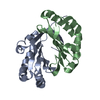
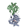
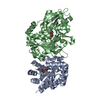
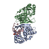
| Title | Structure of the effector-binding domain of deoxyribonucleoside regulator DeoR from Bacillus subtilis in complex with deoxyribose-5-phosphate | ||||||
 Components Components | Deoxyribonucleoside regulator | ||||||
 Keywords Keywords | TRANSCRIPTION / Rossmann Fold / Sugar-binding transcriptional regulator / Schiff base | ||||||
| Function / homology |  Function and homology information Function and homology informationregulation of DNA-templated transcription initiation / cis-regulatory region sequence-specific DNA binding / carbohydrate binding / identical protein binding Similarity search - Function | ||||||
| Biological species |  | ||||||
| Method |  X-RAY DIFFRACTION / SYNCHROTRON / MOLECULAR REPLACEMENT / Resolution: 1.6 Å X-RAY DIFFRACTION / SYNCHROTRON / MOLECULAR REPLACEMENT / Resolution: 1.6 Å | ||||||
 Authors Authors | Rezacova, P. / Skerlova, J. | ||||||
 Citation Citation | Journal: Febs J. / Year: 2014 Title: Structure of the effector-binding domain of deoxyribonucleoside regulator DeoR from Bacillus subtilis. Authors: Skerlova, J. / Fabry, M. / Hubalek, M. / Otwinowski, Z. / Rezacova, P. #1: Journal: Mol.Microbiol. / Year: 2008Title: Crystal structures of the effector-binding domain of repressor Central glycolytic gene Regulator from Bacillus subtilis reveal ligand-induced structural changes upon binding of several glycolytic intermediates. Authors: Rezacova, P. / Kozisek, M. / Moy, S.F. / Sieglova, I. / Joachimiak, A. / Machius, M. / Otwinowski, Z. #2: Journal: Cryst.GrowthDes. / Year: 2013Title: Crystallization of the Effector-Binding Domain of Repressor DeoR from Bacillus subtilis Authors: Pisackova, J. / Prochazkova, K. / Fabry, M. / Rezacova, P. | ||||||
| History |
|
- Structure visualization
| Structure viewer | Molecule: MolmilJmol/JSmol |
|---|
- Downloads & links
-Download
| PDBx/mmCIF format | 4oqp.cif.gz | 128.1 KB | Display | PDBx/mmCIF format |
|---|---|---|---|---|
| PDB format | pdb4oqp.ent.gz | 99.1 KB | Display | PDB format |
| PDBx/mmJSON format | 4oqp.json.gz | Tree view | PDBx/mmJSON format | |
| Others |  Other downloads Other downloads |
-Validation report
| Arichive directory | https://data.pdbj.org/pub/pdb/validation_reports/oq/4oqpftp://data.pdbj.org/pub/pdb/validation_reports/oq/4oqp | HTTPS FTP |
|---|
-Related structure data
| Related structure data | 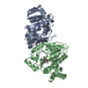 4oqqC  3nzeS C: citing same article ( S: Starting model for refinement |
|---|---|
| Similar structure data |


-Links
 PDBj
PDBj

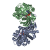
- Assembly
| Deposited unit | 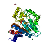
| |||||||||||||||
|---|---|---|---|---|---|---|---|---|---|---|---|---|---|---|---|---|
| 1 | 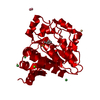
| |||||||||||||||
| Unit cell |
| |||||||||||||||
| Components on special symmetry positions |
|
-Components
-Protein , 1 types, 1 molecules A
| #1: Protein | Mass: 29371.447 Da / Num. of mol.: 1 / Fragment: C-terminal domain Source method: isolated from a genetically manipulated source Source: (gene. exp.) Strain: 168 / Gene: deoR, yxxC, BSU39430 / Plasmid: pET151 / Production host: |
|---|
-Non-polymers , 6 types, 271 molecules 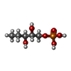


| #2: Chemical | ChemComp-PED /  Mass: 200.127 Da / Num. of mol.: 1 / Source method: obtained synthetically / Formula: C5H13O6P Mass: 200.127 Da / Num. of mol.: 1 / Source method: obtained synthetically / Formula: C5H13O6P | ||||||
|---|---|---|---|---|---|---|---|
| #3: Chemical | ChemComp-CD /  Mass: 112.411 Da / Num. of mol.: 1 / Source method: obtained synthetically / Formula: Cd Mass: 112.411 Da / Num. of mol.: 1 / Source method: obtained synthetically / Formula: Cd | ||||||
| #4: Chemical |  Mass: 58.693 Da / Num. of mol.: 3 / Source method: obtained synthetically / Formula: Ni Mass: 58.693 Da / Num. of mol.: 3 / Source method: obtained synthetically / Formula: Ni#5: Chemical | ChemComp-CO / |  Mass: 58.933 Da / Num. of mol.: 1 / Source method: obtained synthetically / Formula: Co Mass: 58.933 Da / Num. of mol.: 1 / Source method: obtained synthetically / Formula: Co#6: Chemical | ChemComp-MG / |  Mass: 24.305 Da / Num. of mol.: 1 / Source method: obtained synthetically / Formula: Mg Mass: 24.305 Da / Num. of mol.: 1 / Source method: obtained synthetically / Formula: Mg#7: Water | ChemComp-HOH / | Mass: 18.015 Da / Num. of mol.: 264 / Source method: isolated from a natural source / Formula: H2O |
-Details
| Has protein modification | Y |
|---|---|
| Nonpolymer details | THE ORGINAL SUGAR D 2-DEOXYRIBOSE-5-PHOSPHATE WAS COVALENTLY BOUND TO LYS141 BY SCHIFF BASE. THE ...THE ORGINAL SUGAR D 2-DEOXYRIBOS |
-Experimental details
-Experiment
| Experiment | Method: X-RAY DIFFRACTION / Number of used crystals: 1 |
|---|
- Sample preparation
| Crystal | Density Matthews: 2.36 Å3/Da / Density % sol: 47.97 % |
|---|---|
| Crystal grow | Temperature: 291 K / pH: 7.5 Details: 0.08 M HEPES, 9.6% (w/v) PEG 3350, 4 mM CoCl2, 4 mM CdCl2, 4 mM MgCl2, and 4 mM NiCl2; 16.5 mg/mL protein in 20 mM trisodium citrate pH 7.0, 150 mM NaCl, 0.02% (v/v) 2-mercaptoethanol, 50 mM ...Details: 0.08 M HEPES, 9.6% (w/v) PEG 3350, 4 mM CoCl2, 4 mM CdCl2, 4 mM MgCl2, and 4 mM NiCl2; 16.5 mg/mL protein in 20 mM trisodium citrate pH 7.0, 150 mM NaCl, 0.02% (v/v) 2-mercaptoethanol, 50 mM deoxyribose-5-phosphate; protein:reservoir 2:1, VAPOR DIFFUSION, HANGING DROP, temperature 291K |
-Data collection
| Diffraction | Mean temperature: 100 K |
|---|---|
| Diffraction source | Source: SYNCHROTRON / Site: BESSY  / Beamline: 14.2 / Wavelength: 0.91841 / Beamline: 14.2 / Wavelength: 0.91841 |
| Detector | Type: MARMOSAIC 225 mm CCD / Detector: CCD / Date: Jul 17, 2012 Details: DOUBLE CRYSTAL MONOCHROMATOR WITH 2 SETS OF RH-SOATED SILLICON AND GLASS MIRRORS |
| Radiation | Monochromator: SI(111) CRYSTAL / Protocol: SINGLE WAVELENGTH / Monochromatic (M) / Laue (L): M / Scattering type: x-ray |
| Radiation wavelength | Wavelength: 0.91841 Å / Relative weight: 1 |
| Reflection | Resolution: 1.6→50 Å / Num. obs: 34660 / % possible obs: 93.5 % / Observed criterion σ(I): 0 / Redundancy: 5.6 % / Rmerge(I) obs: 0.04 |
| Reflection shell | Resolution: 1.6→1.66 Å / Redundancy: 3.3 % / Rmerge(I) obs: 0.255 / Mean I/σ(I) obs: 2.8 / % possible all: 62.3 |
- Processing
| Software |
| ||||||||||||||||||||||||||||||||||||||||||||||||||||||||||||||||||||||||||||||||||||||||||||||||||||||||||||||||||||||||||||||||||||||||||||||||||||||||||||||||||||||||||||||||||||||
|---|---|---|---|---|---|---|---|---|---|---|---|---|---|---|---|---|---|---|---|---|---|---|---|---|---|---|---|---|---|---|---|---|---|---|---|---|---|---|---|---|---|---|---|---|---|---|---|---|---|---|---|---|---|---|---|---|---|---|---|---|---|---|---|---|---|---|---|---|---|---|---|---|---|---|---|---|---|---|---|---|---|---|---|---|---|---|---|---|---|---|---|---|---|---|---|---|---|---|---|---|---|---|---|---|---|---|---|---|---|---|---|---|---|---|---|---|---|---|---|---|---|---|---|---|---|---|---|---|---|---|---|---|---|---|---|---|---|---|---|---|---|---|---|---|---|---|---|---|---|---|---|---|---|---|---|---|---|---|---|---|---|---|---|---|---|---|---|---|---|---|---|---|---|---|---|---|---|---|---|---|---|---|---|
| Refinement | Method to determine structure: MOLECULAR REPLACEMENT Starting model: PDB ENTRY 3NZE Resolution: 1.6→19.77 Å / Cor.coef. Fo:Fc: 0.972 / Cor.coef. Fo:Fc free: 0.958 / SU B: 2.784 / SU ML: 0.055 / Cross valid method: THROUGHOUT / σ(F): 0 / ESU R: 0.082 / ESU R Free: 0.085 / Stereochemistry target values: MAXIMUM LIKELIHOOD / Details: HYDROGENS HAVE BEEN ADDED IN THE RIDING POSITIONS
| ||||||||||||||||||||||||||||||||||||||||||||||||||||||||||||||||||||||||||||||||||||||||||||||||||||||||||||||||||||||||||||||||||||||||||||||||||||||||||||||||||||||||||||||||||||||
| Solvent computation | Ion probe radii: 0.8 Å / Shrinkage radii: 0.8 Å / VDW probe radii: 1.2 Å / Solvent model: BABINET MODEL WITH MASK | ||||||||||||||||||||||||||||||||||||||||||||||||||||||||||||||||||||||||||||||||||||||||||||||||||||||||||||||||||||||||||||||||||||||||||||||||||||||||||||||||||||||||||||||||||||||
| Displacement parameters | Biso mean: 24.6 Å2
| ||||||||||||||||||||||||||||||||||||||||||||||||||||||||||||||||||||||||||||||||||||||||||||||||||||||||||||||||||||||||||||||||||||||||||||||||||||||||||||||||||||||||||||||||||||||
| Refinement step | Cycle: LAST / Resolution: 1.6→19.77 Å
| ||||||||||||||||||||||||||||||||||||||||||||||||||||||||||||||||||||||||||||||||||||||||||||||||||||||||||||||||||||||||||||||||||||||||||||||||||||||||||||||||||||||||||||||||||||||
| Refine LS restraints |
|