 Movie
Movie Controller

[English] 日本語
 Yorodumi
- PDB-4okd: Crystal Structure of Chlamydomonas reinhardtii Isoamylase 1 (ISA1... -
+ Open data

- Basic information
| Entry | Database: PDB / ID: 4okd | |||||||||
|---|---|---|---|---|---|---|---|---|---|---|
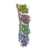
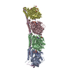
| Title | Crystal Structure of Chlamydomonas reinhardtii Isoamylase 1 (ISA1) in complex with maltoheptaose | |||||||||
 Components Components | Isoamylase | |||||||||
 Keywords Keywords | HYDROLASE / GH13 glycoside hydrolase | |||||||||
| Function / homology |  Function and homology information Function and homology informationisoamylase / isoamylase activity / chloroplast / carbohydrate metabolic process Similarity search - Function | |||||||||
| Biological species |   Chlamydomonas reinhardtii (plant) Chlamydomonas reinhardtii (plant) | |||||||||
| Method |  X-RAY DIFFRACTION / SYNCHROTRON / MOLECULAR REPLACEMENT / Resolution: 2.4 Å X-RAY DIFFRACTION / SYNCHROTRON / MOLECULAR REPLACEMENT / Resolution: 2.4 Å | |||||||||
 Authors Authors | Sim, L. / Palcic, M. | |||||||||
 Citation Citation | Journal: J.Biol.Chem. / Year: 2014 Title: Crystal Structure of the Chlamydomonas Starch Debranching Enzyme Isoamylase ISA1 Reveals Insights into the Mechanism of Branch Trimming and Complex Assembly. Authors: Sim, L. / Beeren, S.R. / Findinier, J. / Dauvillee, D. / Ball, S.G. / Henriksen, A. / Palcic, M.M. | |||||||||
| History |
|
- Structure visualization
| Structure viewer | Molecule: MolmilJmol/JSmol |
|---|
- Downloads & links
-Download
| PDBx/mmCIF format | 4okd.cif.gz | 638.6 KB | Display | PDBx/mmCIF format |
|---|---|---|---|---|
| PDB format | pdb4okd.ent.gz | 526.5 KB | Display | PDB format |
| PDBx/mmJSON format | 4okd.json.gz | Tree view | PDBx/mmJSON format | |
| Others |  Other downloads Other downloads |
-Validation report
| Arichive directory | https://data.pdbj.org/pub/pdb/validation_reports/ok/4okdftp://data.pdbj.org/pub/pdb/validation_reports/ok/4okd | HTTPS FTP |
|---|
-Related structure data
| Related structure data |  4j7rC  4jr7S S: Starting model for refinement C: citing same article ( |
|---|---|
| Similar structure data | |
| Other databases |
|


-Links
 PDBj
PDBj


- Assembly
| Deposited unit | 
| ||||||||
|---|---|---|---|---|---|---|---|---|---|
| 1 |
| ||||||||
| Unit cell |
|
-Components
| #1: Protein | Mass: 93054.656 Da / Num. of mol.: 2 / Fragment: UNP Residues 57-875 Source method: isolated from a genetically manipulated source Source: (gene. exp.) Chlamydomonas reinhardtii (plant) / Gene: ISA1 / Plasmid: pET28a / Production host:  #2: Polysaccharide | Source method: isolated from a genetically manipulated source #3: Polysaccharide |   Source method: isolated from a genetically manipulated source Details: oligosaccharide / References: alpha-maltotriose #4: Sugar | ChemComp-GLC / |   Type: D-saccharide, alpha linking / Mass: 180.156 Da / Num. of mol.: 1 Type: D-saccharide, alpha linking / Mass: 180.156 Da / Num. of mol.: 1Source method: isolated from a genetically manipulated source Formula: C6H12O6 #5: Water | ChemComp-HOH / |  Mass: 18.015 Da / Num. of mol.: 433 / Source method: isolated from a natural source / Formula: H2O Mass: 18.015 Da / Num. of mol.: 433 / Source method: isolated from a natural source / Formula: H2OHas protein modification | Y | |
|---|
-Experimental details
-Experiment
| Experiment | Method: X-RAY DIFFRACTION / Number of used crystals: 1 |
|---|
- Sample preparation
| Crystal | Density Matthews: 3.46 Å3/Da / Density % sol: 64.44 % |
|---|---|
| Crystal grow | Temperature: 293 K / pH: 7 Details: 0.2M ammonium citrate tribasic, 14-20% PEG3350, pH 7.0, vapor diffusion, hanging drop, temperature 293K |
-Data collection
| Diffraction | Mean temperature: 100 K |
|---|---|
| Diffraction source | Source: SYNCHROTRON / Site: ESRF  / Beamline: ID23-2 / Wavelength: 0.8726 / Beamline: ID23-2 / Wavelength: 0.8726 |
| Detector | Type: MARMOSAIC 225 mm CCD / Detector: CCD / Date: Sep 15, 2012 |
| Radiation | Protocol: SINGLE WAVELENGTH / Monochromatic (M) / Laue (L): M / Scattering type: x-ray |
| Radiation wavelength | Wavelength: 0.8726 Å / Relative weight: 1 |
| Reflection | Resolution: 2.3→50 Å / Num. obs: 109429 / % possible obs: 93 % / Observed criterion σ(I): -3 / Biso Wilson estimate: 51.81 Å2 / Rmerge(I) obs: 0.118 / Net I/σ(I): 13.33 |
| Reflection shell | Resolution: 2.3→2.44 Å / Rmerge(I) obs: 0.014 / Mean I/σ(I) obs: 1.37 / % possible all: 64.5 |
- Processing
| Software |
| ||||||||||||||||||||||||||||||||||||||||||||||||||||||||||||||||||||||||||||||||||||||||||||||||||||||||||||||||||||||||||||||||||||||||||||||||||||||||||||||||||||||||||||||||||||||
|---|---|---|---|---|---|---|---|---|---|---|---|---|---|---|---|---|---|---|---|---|---|---|---|---|---|---|---|---|---|---|---|---|---|---|---|---|---|---|---|---|---|---|---|---|---|---|---|---|---|---|---|---|---|---|---|---|---|---|---|---|---|---|---|---|---|---|---|---|---|---|---|---|---|---|---|---|---|---|---|---|---|---|---|---|---|---|---|---|---|---|---|---|---|---|---|---|---|---|---|---|---|---|---|---|---|---|---|---|---|---|---|---|---|---|---|---|---|---|---|---|---|---|---|---|---|---|---|---|---|---|---|---|---|---|---|---|---|---|---|---|---|---|---|---|---|---|---|---|---|---|---|---|---|---|---|---|---|---|---|---|---|---|---|---|---|---|---|---|---|---|---|---|---|---|---|---|---|---|---|---|---|---|---|
| Refinement | Method to determine structure: MOLECULAR REPLACEMENT Starting model: PDB ENTRY 4JR7 Resolution: 2.4→45.95 Å / Cor.coef. Fo:Fc: 0.955 / Cor.coef. Fo:Fc free: 0.938 / SU B: 15.544 / SU ML: 0.168 / SU R Cruickshank DPI: 0.2582 / Cross valid method: THROUGHOUT / σ(F): 0 / ESU R: 0.258 / ESU R Free: 0.206 / Stereochemistry target values: MAXIMUM LIKELIHOOD / Details: HYDROGENS HAVE BEEN ADDED IN THE RIDING
| ||||||||||||||||||||||||||||||||||||||||||||||||||||||||||||||||||||||||||||||||||||||||||||||||||||||||||||||||||||||||||||||||||||||||||||||||||||||||||||||||||||||||||||||||||||||
| Solvent computation | Ion probe radii: 0.8 Å / Shrinkage radii: 0.8 Å / VDW probe radii: 1.2 Å / Solvent model: MASK | ||||||||||||||||||||||||||||||||||||||||||||||||||||||||||||||||||||||||||||||||||||||||||||||||||||||||||||||||||||||||||||||||||||||||||||||||||||||||||||||||||||||||||||||||||||||
| Displacement parameters | Biso mean: 51.92 Å2
| ||||||||||||||||||||||||||||||||||||||||||||||||||||||||||||||||||||||||||||||||||||||||||||||||||||||||||||||||||||||||||||||||||||||||||||||||||||||||||||||||||||||||||||||||||||||
| Refinement step | Cycle: LAST / Resolution: 2.4→45.95 Å
| ||||||||||||||||||||||||||||||||||||||||||||||||||||||||||||||||||||||||||||||||||||||||||||||||||||||||||||||||||||||||||||||||||||||||||||||||||||||||||||||||||||||||||||||||||||||
| Refine LS restraints |
|