 Movie
Movie Controller

[English] 日本語
 Yorodumi
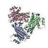
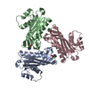
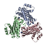
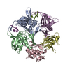
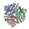
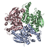
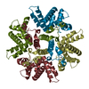
- PDB-4ok9: Crystal structure of the single-stranded RNA binding protein HutP... -
+ Open data

- Basic information
| Entry | Database: PDB / ID: 4ok9 | ||||||
|---|---|---|---|---|---|---|---|
| Title | Crystal structure of the single-stranded RNA binding protein HutP from Geobacillus thermodenitrificans | ||||||
 Components Components | Hut operon positive regulatory protein | ||||||
 Keywords Keywords | RNA BINDING PROTEIN / Antitermination / Single stranded RNA binding protein | ||||||
| Function / homology |  Function and homology information Function and homology information: / mRNA binding / positive regulation of gene expression / DNA-templated transcription Similarity search - Function | ||||||
| Biological species |  Geobacillus thermodenitrificans (bacteria) Geobacillus thermodenitrificans (bacteria) | ||||||
| Method |  X-RAY DIFFRACTION / SYNCHROTRON / MOLECULAR REPLACEMENT / Resolution: 1.91 Å X-RAY DIFFRACTION / SYNCHROTRON / MOLECULAR REPLACEMENT / Resolution: 1.91 Å | ||||||
 Authors Authors | Thiruselvam, V. / Ponnuswamy, M.N. / Kumarevel, T.S. | ||||||
 Citation Citation | Journal: Biochem.Biophys.Res.Commun. / Year: 2014 Title: Crystal structure of the single-stranded RNA binding protein HutP from Geobacillus thermodenitrificans Authors: Thiruselvam, V. / Sivaraman, P. / Kumarevel, T. / Ponnuswamy, M.N. | ||||||
| History |
|
- Structure visualization
| Structure viewer | Molecule: MolmilJmol/JSmol |
|---|
- Downloads & links
-Download
| PDBx/mmCIF format | 4ok9.cif.gz | 73.8 KB | Display | PDBx/mmCIF format |
|---|---|---|---|---|
| PDB format | pdb4ok9.ent.gz | 55.4 KB | Display | PDB format |
| PDBx/mmJSON format | 4ok9.json.gz | Tree view | PDBx/mmJSON format | |
| Others |  Other downloads Other downloads |
-Validation report
| Arichive directory | https://data.pdbj.org/pub/pdb/validation_reports/ok/4ok9ftp://data.pdbj.org/pub/pdb/validation_reports/ok/4ok9 | HTTPS FTP |
|---|
-Related structure data
| Related structure data | 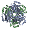 4okqC  2zh0 C: citing same article ( S: Starting model for refinement |
|---|---|
| Similar structure data |


-Links
 PDBj
PDBj
- Assembly
| Deposited unit | 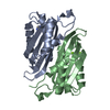
| ||||||||||||||||||||||||
|---|---|---|---|---|---|---|---|---|---|---|---|---|---|---|---|---|---|---|---|---|---|---|---|---|---|
| 1 | 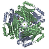
| ||||||||||||||||||||||||
| Unit cell |
| ||||||||||||||||||||||||
| Components on special symmetry positions |
|
-Components
| #1: Protein | Mass: 16302.699 Da / Num. of mol.: 2 / Fragment: HutP Source method: isolated from a genetically manipulated source Source: (gene. exp.) Geobacillus thermodenitrificans (bacteria)Strain: NG80-2 / Gene: GTNG_0361, hutP / Production host: #2: Chemical | ChemComp-MG /   Mass: 24.305 Da / Num. of mol.: 6 / Source method: obtained synthetically / Formula: Mg Mass: 24.305 Da / Num. of mol.: 6 / Source method: obtained synthetically / Formula: Mg#3: Chemical |   Type: L-peptide linking / Mass: 156.162 Da / Num. of mol.: 2 / Source method: obtained synthetically / Formula: C6H10N3O2 Type: L-peptide linking / Mass: 156.162 Da / Num. of mol.: 2 / Source method: obtained synthetically / Formula: C6H10N3O2#4: Water | ChemComp-HOH / |  Mass: 18.015 Da / Num. of mol.: 181 / Source method: isolated from a natural source / Formula: H2O Mass: 18.015 Da / Num. of mol.: 181 / Source method: isolated from a natural source / Formula: H2O |
|---|
-Experimental details
-Experiment
| Experiment | Method: X-RAY DIFFRACTION / Number of used crystals: 1 |
|---|
- Sample preparation
| Crystal | Density Matthews: 2.8 Å3/Da / Density % sol: 56.06 % Description: THE ENTRY CONTAINS FRIEDEL PAIRS IN F_PLUS/MINUS COLUMNS. |
|---|---|
| Crystal grow | Temperature: 293 K / Method: vapor diffusion, hanging drop / pH: 9.5 Details: 10%(W/V) PEG300, 0.1M CHES, pH 9.5, VAPOR DIFFUSION, HANGING DROP, temperature 293K |
-Data collection
| Diffraction | Mean temperature: 100 K |
|---|---|
| Diffraction source | Source: SYNCHROTRON / Site: SPring-8  / Beamline: BL26B1 / Wavelength: 1 Å / Beamline: BL26B1 / Wavelength: 1 Å |
| Detector | Type: RIGAKU SATURN A200 / Detector: CCD / Date: Jan 23, 2013 / Details: Si II crystal |
| Radiation | Monochromator: Si double crystal monochromator / Protocol: SINGLE WAVELENGTH / Monochromatic (M) / Laue (L): M / Scattering type: x-ray |
| Radiation wavelength | Wavelength: 1 Å / Relative weight: 1 |
| Reflection | Resolution: 1.91→50 Å / Num. all: 27972 / Num. obs: 27972 / % possible obs: 99.9 % / Observed criterion σ(F): 2 / Observed criterion σ(I): 2 / Redundancy: 16 % / Rmerge(I) obs: 0.071 / Net I/σ(I): 56 |
| Reflection shell | Resolution: 1.91→1.94 Å / Redundancy: 14.5 % / Rmerge(I) obs: 0.449 / Mean I/σ(I) obs: 5.2 / Num. unique all: 1388 / % possible all: 100 |
- Processing
| Software |
| ||||||||||||||||||||||||||||||||||||||||||||||||||||||||||||
|---|---|---|---|---|---|---|---|---|---|---|---|---|---|---|---|---|---|---|---|---|---|---|---|---|---|---|---|---|---|---|---|---|---|---|---|---|---|---|---|---|---|---|---|---|---|---|---|---|---|---|---|---|---|---|---|---|---|---|---|---|---|
| Refinement | Method to determine structure: MOLECULAR REPLACEMENT Starting model: 2ZH0 2zh0 Resolution: 1.91→35.01 Å / Cor.coef. Fo:Fc: 0.967 / Cor.coef. Fo:Fc free: 0.936 / SU B: 3.315 / SU ML: 0.096 / Cross valid method: THROUGHOUT / σ(F): 3 / ESU R: 0.131 / ESU R Free: 0.137 / Stereochemistry target values: MAXIMUM LIKELIHOOD Details: HYDROGENS HAVE BEEN ADDED IN THE RIDING POSITIONS, SF FILE CONTAINS FRIEDEL PAIRS UNDER I/F_MINUS AND I/F_PLUS COLUMNS.
| ||||||||||||||||||||||||||||||||||||||||||||||||||||||||||||
| Solvent computation | Ion probe radii: 0.8 Å / Shrinkage radii: 0.8 Å / VDW probe radii: 1.2 Å / Solvent model: MASK | ||||||||||||||||||||||||||||||||||||||||||||||||||||||||||||
| Displacement parameters | Biso mean: 35.348 Å2
| ||||||||||||||||||||||||||||||||||||||||||||||||||||||||||||
| Refinement step | Cycle: LAST / Resolution: 1.91→35.01 Å
| ||||||||||||||||||||||||||||||||||||||||||||||||||||||||||||
| Refine LS restraints |
| ||||||||||||||||||||||||||||||||||||||||||||||||||||||||||||
| LS refinement shell | Resolution: 1.909→1.959 Å / Total num. of bins used: 20
|
