Mass: 18.015 Da / Num. of mol.: 542 / Source method: isolated from a natural source / Formula: H2O
Has protein modification
Y
-
Experimental details
-
Experiment
Experiment
Method: X-RAY DIFFRACTION / Number of used crystals: 1
-
Sample preparation
Crystal
Density Matthews: 2.26 Å3/Da / Density % sol: 45.49 %
Crystal grow
Temperature: 293 K / Method: vapor diffusion, hanging drop Details: 22% PEG3350, 0.2 M KSCN, 0.2 M NDSB-221, VAPOR DIFFUSION, HANGING DROP, temperature 293K
-
Data collection
Diffraction
Mean temperature: 100 K
Diffraction source
Source: SYNCHROTRON / Site: Australian Synchrotron / Beamline: MX2 / Wavelength: 0.9537 Å
Resolution: 1.597→37.718 Å / σ(F): 1.96 / Phase error: 22.46 / Stereochemistry target values: TWIN_LSQ_F Details: the following twin law was applied in the refinement: h,-h-k,-l
Rfactor
Num. reflection
% reflection
Selection details
Rfree
0.1995
3774
5.03 %
RANDOM
Rwork
0.1725
-
-
-
all
0.178
74969
-
-
obs
0.178
74969
98.61 %
-
Solvent computation
Shrinkage radii: 0.9 Å / VDW probe radii: 1.11 Å / Solvent model: FLAT BULK SOLVENT MODEL
Refinement step
Cycle: LAST / Resolution: 1.597→37.718 Å
Protein
Nucleic acid
Ligand
Solvent
Total
Num. atoms
4512
0
12
542
5066
Refine LS restraints
Refine-ID
Type
Dev ideal
Number
X-RAY DIFFRACTION
f_bond_d
0.017
4620
X-RAY DIFFRACTION
f_angle_d
1.555
6256
X-RAY DIFFRACTION
f_dihedral_angle_d
14.482
1666
X-RAY DIFFRACTION
f_chiral_restr
0.071
696
X-RAY DIFFRACTION
f_plane_restr
0.009
814
LS refinement shell
Resolution (Å)
Rfactor Rfree
Num. reflection Rfree
Rfactor Rwork
Num. reflection Rwork
Refine-ID
% reflection obs (%)
1.5982-1.6257
0.2928
182
0.3037
3221
X-RAY DIFFRACTION
85
1.6257-1.6553
0.2855
191
0.2777
3594
X-RAY DIFFRACTION
93
1.6553-1.6871
0.2941
170
0.2697
3563
X-RAY DIFFRACTION
94
1.6871-1.7215
0.2847
194
0.2772
3547
X-RAY DIFFRACTION
94
1.7215-1.759
0.3056
196
0.2557
3545
X-RAY DIFFRACTION
94
1.759-1.7999
0.2854
154
0.2592
3577
X-RAY DIFFRACTION
95
1.7999-1.8449
0.2573
192
0.2376
3560
X-RAY DIFFRACTION
94
1.8449-1.8947
0.2489
169
0.2278
3599
X-RAY DIFFRACTION
95
1.8947-1.9504
0.2337
192
0.2221
3581
X-RAY DIFFRACTION
94
1.9504-2.0134
0.2388
187
0.2204
3516
X-RAY DIFFRACTION
94
2.0134-2.0853
0.23
205
0.2127
3608
X-RAY DIFFRACTION
94
2.0853-2.1687
0.2511
176
0.2029
3590
X-RAY DIFFRACTION
95
2.1687-2.2674
0.2233
206
0.2002
3535
X-RAY DIFFRACTION
94
2.2674-2.3868
0.2264
206
0.1929
3611
X-RAY DIFFRACTION
94
2.3868-2.5362
0.202
182
0.1877
3554
X-RAY DIFFRACTION
95
2.5362-2.7317
0.2073
188
0.1767
3621
X-RAY DIFFRACTION
95
2.7317-3.0061
0.2029
189
0.1667
3586
X-RAY DIFFRACTION
95
3.0061-3.4399
0.1948
194
0.1506
3583
X-RAY DIFFRACTION
95
3.4399-4.3295
0.1486
199
0.1218
3580
X-RAY DIFFRACTION
95
4.3295-23.4636
0.1508
193
0.1219
3592
X-RAY DIFFRACTION
95
Refinement TLS params.
Method: refined / Refine-ID: X-RAY DIFFRACTION
ID
L11 (°2)
L12 (°2)
L13 (°2)
L22 (°2)
L23 (°2)
L33 (°2)
S11 (Å °)
S12 (Å °)
S13 (Å °)
S21 (Å °)
S22 (Å °)
S23 (Å °)
S31 (Å °)
S32 (Å °)
S33 (Å °)
T11 (Å2)
T12 (Å2)
T13 (Å2)
T22 (Å2)
T23 (Å2)
T33 (Å2)
Origin x (Å)
Origin y (Å)
Origin z (Å)
1
0.829
0.1255
-0.0435
1.5344
-1.1797
3.7327
-0.0194
-0.0395
0.1029
0.0529
-0.0298
0.0267
-0.5942
-0.1875
0.0514
0.2185
0.0657
-0.0145
0.1119
-0.0234
0.1147
-34.7841
24.8217
22.4453
2
2.1838
1.0592
-0.8289
1.3407
-0.6469
0.9605
-0.0716
0.1533
-0.0044
-0.1213
0.1093
0.0889
0.0639
-0.0386
-0.0431
0.1584
-0.0041
-0.0114
0.1319
-0.0179
0.1385
0.306
37.9563
3.5805
3
1.0243
0.3483
1.1057
1.1303
0.437
3.7485
-0.0225
0.1501
0.0203
-0.0412
-0.0163
-0.1055
-0.1095
0.5884
0.0369
0.0793
0.0035
0.0182
0.2447
0.0105
0.1185
1.9306
3.6445
13.9395
4
9.2741
6.4686
-0.8967
7.6135
-1.8552
4.1555
-0.0249
-0.3712
0.4005
0.2399
-0.0025
0.4446
-0.5237
-0.1467
0.0346
0.2271
0.0677
-0.0291
0.1055
-0.0078
0.1354
-37.1989
29.7927
9.0588
5
4.9572
1.7858
1.7065
3.4231
2.781
3.5114
0.1805
0.0378
-0.0241
0.0862
-0.0778
-0.3164
0.1011
0.5931
-0.058
0.1421
0.0049
0.0189
0.3061
0.0438
0.1117
7.3139
4.0021
27.3955
6
1.1246
2.1325
-0.7503
2.0035
-2.5282
1.8731
-0.1049
0.097
-0.0262
-0.6052
0.2711
-0.2242
0.0644
0.0117
-0.1448
0.19
0.01
0.0215
0.2225
-0.0422
0.1725
-5.2907
35.6634
17.0281
Refinement TLS group
ID
Refine-ID
Refine TLS-ID
Selection details
1
X-RAY DIFFRACTION
1
chain 'A'
2
X-RAY DIFFRACTION
2
chain 'B'
3
X-RAY DIFFRACTION
3
chain 'C'
4
X-RAY DIFFRACTION
4
chain 'D'
5
X-RAY DIFFRACTION
5
chain 'E'
6
X-RAY DIFFRACTION
6
chain 'F'
+
About Yorodumi
-
News
-
Feb 9, 2022. New format data for meta-information of EMDB entries
New format data for meta-information of EMDB entries
Version 3 of the EMDB header file is now the official format.
The previous official version 1.9 will be removed from the archive.
In the structure databanks used in Yorodumi, some data are registered as the other names, "COVID-19 virus" and "2019-nCoV". Here are the details of the virus and the list of structure data.
Jan 31, 2019. EMDB accession codes are about to change! (news from PDBe EMDB page)
EMDB accession codes are about to change! (news from PDBe EMDB page)
The allocation of 4 digits for EMDB accession codes will soon come to an end. Whilst these codes will remain in use, new EMDB accession codes will include an additional digit and will expand incrementally as the available range of codes is exhausted. The current 4-digit format prefixed with “EMD-” (i.e. EMD-XXXX) will advance to a 5-digit format (i.e. EMD-XXXXX), and so on. It is currently estimated that the 4-digit codes will be depleted around Spring 2019, at which point the 5-digit format will come into force.
The EM Navigator/Yorodumi systems omit the EMD- prefix.
Related info.:Q: What is EMD? / ID/Accession-code notation in Yorodumi/EM Navigator
Yorodumi is a browser for structure data from EMDB, PDB, SASBDB, etc.
This page is also the successor to EM Navigator detail page, and also detail information page/front-end page for Omokage search.
The word "yorodu" (or yorozu) is an old Japanese word meaning "ten thousand". "mi" (miru) is to see.
Related info.:EMDB / PDB / SASBDB / Comparison of 3 databanks / Yorodumi Search / Aug 31, 2016. New EM Navigator & Yorodumi / Yorodumi Papers / Jmol/JSmol / Function and homology information / Changes in new EM Navigator and Yorodumi
 Movie
Movie Controller

 Yorodumi
 Open data
 Basic information
 Components
 Keywords
 Function and homology information
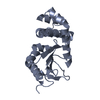
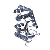
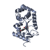
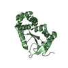
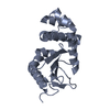
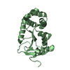
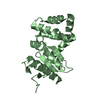
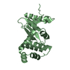
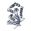
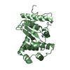
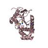
 Proteus mirabilis (bacteria)
 X-RAY DIFFRACTION /
  Authors
 Citation
 Structure visualization
 Downloads & links
 Other downloads


 PDBj
PDBj


 Assembly
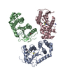

 Mass: 58.082 Da / Num. of mol.: 4 / Source method: obtained synthetically / Formula: CNS
 Mass: 18.015 Da / Num. of mol.: 542 / Source method: isolated from a natural source / Formula: H2O
 Sample preparation
 / Beamline: MX2 / Wavelength: 0.9537 Å
 Processing