| 登録情報 | データベース: PDB / ID: 4oc8
|
|---|
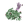
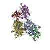
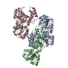
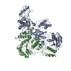
| タイトル | DNA modification-dependent restriction endonuclease AspBHI |
|---|
 要素 要素 | restriction endonuclease AspBHI |
|---|
 キーワード キーワード | HYDROLASE / DNA Cleavage / DNA Restriction Enzymes / DNA-Binding Proteins / Tetramerization / Models / Molecular / Azoarcus / Protein Multimerization / Protein Structure / Tertiary |
|---|
| 機能・相同性 |  機能・相同性情報 機能・相同性情報
DNA restriction-modification system / endonuclease activity / DNA binding類似検索 - 分子機能 PUA domain-like - #20 / Restriction endonuclease AspBHI, N-terminal / Restriction endonuclease AspBHI N-terminal / Restriction endonuclease type IV, Mrr / Restriction endonuclease / PUA domain-like / Trna Endonuclease; Chain: A, domain 1 - #10 / Trna Endonuclease; Chain: A, domain 1 / tRNA endonuclease-like domain superfamily / Roll ...PUA domain-like - #20 / Restriction endonuclease AspBHI, N-terminal / Restriction endonuclease AspBHI N-terminal / Restriction endonuclease type IV, Mrr / Restriction endonuclease / PUA domain-like / Trna Endonuclease; Chain: A, domain 1 - #10 / Trna Endonuclease; Chain: A, domain 1 / tRNA endonuclease-like domain superfamily / Roll / 3-Layer(aba) Sandwich / Mainly Beta / Alpha Beta類似検索 - ドメイン・相同性 |
|---|
| 生物種 |  Azoarcus sp. (バクテリア) Azoarcus sp. (バクテリア) |
|---|
| 手法 |  X線回折 / シンクロトロン / AB INITIO PHASING / 解像度: 2.884 Å X線回折 / シンクロトロン / AB INITIO PHASING / 解像度: 2.884 Å |
|---|
 データ登録者 データ登録者 | Horton, J.R. |
|---|
 引用 引用 | ジャーナル: Sci Rep / 年: 2014
タイトル: Structure and mutagenesis of the DNA modification-dependent restriction endonuclease AspBHI.
著者: Horton, J.R. / Nugent, R.L. / Li, A. / Mabuchi, M.Y. / Fomenkov, A. / Cohen-Karni, D. / Griggs, R.M. / Zhang, X. / Wilson, G.G. / Zheng, Y. / Xu, S.Y. / Cheng, X. |
|---|
| 履歴 | | 登録 | 2014年1月8日 | 登録サイト: RCSB / 処理サイト: RCSB |
|---|
| 改定 1.0 | 2014年3月19日 | Provider: repository / タイプ: Initial release |
|---|
| 改定 1.1 | 2024年2月28日 | Group: Data collection / Database references / Derived calculations
カテゴリ: chem_comp_atom / chem_comp_bond ...chem_comp_atom / chem_comp_bond / database_2 / struct_site
Item: _database_2.pdbx_DOI / _database_2.pdbx_database_accession ..._database_2.pdbx_DOI / _database_2.pdbx_database_accession / _struct_site.pdbx_auth_asym_id / _struct_site.pdbx_auth_comp_id / _struct_site.pdbx_auth_seq_id |
|---|
|
|---|
 ムービー
ムービー コントローラー


 データを開く
 基本情報
基本情報 構造の表示
構造の表示 ダウンロードとリンク
 その他のダウンロード


 PDBj
PDBj 集合体
集合体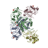

 分子量: 94.971 Da / 分子数: 8 / 由来タイプ: 合成 / 式: PO4
 分子量: 18.015 Da / 分子数: 53 / 由来タイプ: 天然 / 式: H2O
 試料調製
試料調製 / ビームライン: 22-BM / 波長: 1.06296 Å
 解析
解析