 Movie
Movie Controller

[English] 日本語
 Yorodumi
- PDB-4o93: Crystal structure of Thermus thermophilis transhydrogeanse domain... -
+ Open data

- Basic information
| Entry | Database: PDB / ID: 4o93 | ||||||
|---|---|---|---|---|---|---|---|
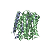
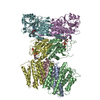
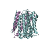
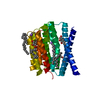
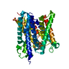
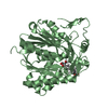
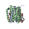
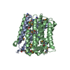
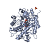
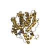
| Title | Crystal structure of Thermus thermophilis transhydrogeanse domain II dimer | ||||||
 Components Components |
| ||||||
 Keywords Keywords | MEMBRANE PROTEIN / Membrane domain dimer | ||||||
| Function / homology |  Function and homology information Function and homology information: / proton-translocating NAD(P)+ transhydrogenase activity / proton-translocating NAD(P)+ transhydrogenase / NADPH regeneration / NADP binding / plasma membrane Similarity search - Function | ||||||
| Biological species |   Thermus thermophilus (bacteria) Thermus thermophilus (bacteria) | ||||||
| Method |  X-RAY DIFFRACTION / SYNCHROTRON / SAD / Resolution: 2.77 Å X-RAY DIFFRACTION / SYNCHROTRON / SAD / Resolution: 2.77 Å | ||||||
 Authors Authors | Leung, J.H. / Yamaguchi, M. / Moeller, A. / Schurig-Briccio, L.A. / Gennis, R.B. / Potter, C.S. / Carragher, B. / Stout, C.D. | ||||||
 Citation Citation | Journal: Science / Year: 2015 Title: Structural biology. Division of labor in transhydrogenase by alternating proton translocation and hydride transfer. Authors: Leung, J.H. / Schurig-Briccio, L.A. / Yamaguchi, M. / Moeller, A. / Speir, J.A. / Gennis, R.B. / Stout, C.D. | ||||||
| History |
|
- Structure visualization
| Structure viewer | Molecule: MolmilJmol/JSmol |
|---|
- Downloads & links
-Download
| PDBx/mmCIF format | 4o93.cif.gz | 137.4 KB | Display | PDBx/mmCIF format |
|---|---|---|---|---|
| PDB format | pdb4o93.ent.gz | 109.3 KB | Display | PDB format |
| PDBx/mmJSON format | 4o93.json.gz | Tree view | PDBx/mmJSON format | |
| Others |  Other downloads Other downloads |
-Validation report
| Summary document | 4o93_validation.pdf.gz | 463 KB | Display | wwPDB validaton report |
|---|---|---|---|---|
| Full document | 4o93_full_validation.pdf.gz | 495.8 KB | Display | |
| Data in XML | 4o93_validation.xml.gz | 28.5 KB | Display | |
| Data in CIF | 4o93_validation.cif.gz | 38.7 KB | Display | |
| Arichive directory | https://data.pdbj.org/pub/pdb/validation_reports/o9/4o93ftp://data.pdbj.org/pub/pdb/validation_reports/o9/4o93 | HTTPS FTP |
-Related structure data


-Links
 PDBj
PDBj


- Assembly
| Deposited unit | 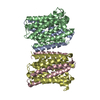
| ||||||||
|---|---|---|---|---|---|---|---|---|---|
| 1 | 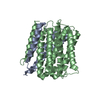
| ||||||||
| 2 | 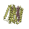
| ||||||||
| 3 |
| ||||||||
| Unit cell |
|
-Components
| #1: Protein | Mass: 10168.106 Da / Num. of mol.: 2 Source method: isolated from a genetically manipulated source Source: (gene. exp.) Thermus thermophilus (bacteria) / Strain: HB27 / ATCC BAA-163 / DSM 7039 / Gene: TT_C1779 / Production host: #2: Protein | Mass: 28109.693 Da / Num. of mol.: 2 Source method: isolated from a genetically manipulated source Source: (gene. exp.) Thermus thermophilus (bacteria) / Strain: HB27 / ATCC BAA-163 / DSM 7039 / Gene: TT_C1778 / Production host: #3: Chemical |   Mass: 200.590 Da / Num. of mol.: 2 / Source method: obtained synthetically / Formula: Hg Mass: 200.590 Da / Num. of mol.: 2 / Source method: obtained synthetically / Formula: Hg#4: Water | ChemComp-HOH / |  Mass: 18.015 Da / Num. of mol.: 9 / Source method: isolated from a natural source / Formula: H2O Mass: 18.015 Da / Num. of mol.: 9 / Source method: isolated from a natural source / Formula: H2O |
|---|
-Experimental details
-Experiment
| Experiment | Method: X-RAY DIFFRACTION / Number of used crystals: 1 |
|---|
- Sample preparation
| Crystal | Density Matthews: 3.51 Å3/Da / Density % sol: 64.98 % |
|---|---|
| Crystal grow | Temperature: 298 K / Method: lipidic subic phase / pH: 8.5 Details: 100 mM Tris pH 8.5, 350 mM NH4-formate, 100mM Na-thiocynate, and 18% (v/v) 1-4-butanediol, lipidic subic phase, temperature 298K |
-Data collection
| Diffraction | Mean temperature: 100 K |
|---|---|
| Diffraction source | Source: SYNCHROTRON / Site: SSRL  / Beamline: BL11-1 / Wavelength: 0.9993 Å / Beamline: BL11-1 / Wavelength: 0.9993 Å |
| Detector | Type: DECTRIS PILATUS 6M / Detector: PIXEL |
| Radiation | Monochromator: Side scattering bent cube-root I-beam single crystal; asymmetric cut 4.965 degs Protocol: SINGLE WAVELENGTH / Monochromatic (M) / Laue (L): M / Scattering type: x-ray |
| Radiation wavelength | Wavelength: 0.9993 Å / Relative weight: 1 |
| Reflection | Resolution: 2.77→98.7 Å / Num. obs: 24865 / Observed criterion σ(F): 0 / Observed criterion σ(I): 0 |
- Processing
| Software |
| |||||||||||||||||||||||||||||||||||||||||||||||||||||||||||||||||
|---|---|---|---|---|---|---|---|---|---|---|---|---|---|---|---|---|---|---|---|---|---|---|---|---|---|---|---|---|---|---|---|---|---|---|---|---|---|---|---|---|---|---|---|---|---|---|---|---|---|---|---|---|---|---|---|---|---|---|---|---|---|---|---|---|---|---|
| Refinement | Method to determine structure: SAD / Resolution: 2.77→98.67 Å / Cor.coef. Fo:Fc: 0.953 / Cor.coef. Fo:Fc free: 0.922 / SU B: 12.749 / SU ML: 0.253 / Cross valid method: THROUGHOUT / ESU R: 0.663 / ESU R Free: 0.351 / Stereochemistry target values: MAXIMUM LIKELIHOOD / Details: HYDROGENS HAVE BEEN USED IF PRESENT IN THE INPUT
| |||||||||||||||||||||||||||||||||||||||||||||||||||||||||||||||||
| Solvent computation | Ion probe radii: 0.8 Å / Shrinkage radii: 0.8 Å / VDW probe radii: 1.2 Å / Solvent model: MASK | |||||||||||||||||||||||||||||||||||||||||||||||||||||||||||||||||
| Displacement parameters | Biso mean: 83.705 Å2
| |||||||||||||||||||||||||||||||||||||||||||||||||||||||||||||||||
| Refinement step | Cycle: LAST / Resolution: 2.77→98.67 Å
| |||||||||||||||||||||||||||||||||||||||||||||||||||||||||||||||||
| Refine LS restraints |
| |||||||||||||||||||||||||||||||||||||||||||||||||||||||||||||||||
| LS refinement shell | Resolution: 2.77→2.842 Å / Total num. of bins used: 20
|
