 Movie
Movie Controller

[English] 日本語
 Yorodumi
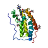
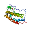
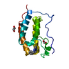
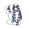
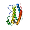
- PDB-4o72: Crystal structure of the first bromodomain of human BRD4 in compl... -
+ Open data

- Basic information
| Entry | Database: PDB / ID: 4o72 | ||||||
|---|---|---|---|---|---|---|---|
| Title | Crystal structure of the first bromodomain of human BRD4 in complex with NU7441 | ||||||
 Components Components | Bromodomain-containing protein 4 | ||||||
 Keywords Keywords | TRANSCRIPTION/INHIBITOR / BROMODOMAIN / CAP / HUNK1 / MCAP / PROTEIN BINDING-INHIBITOR COMPLEX / MITOTIC CHROMOSOME ASSOCIATED PROTEIN / CELL CYCLE / INHIBITOR / TRANSCRIPTION-INHIBITOR complex | ||||||
| Function / homology |  Function and homology information Function and homology informationhistone H4K8ac reader activity / RNA polymerase II C-terminal domain binding / histone H3K27ac reader activity / P-TEFb complex binding / histone H3K9ac reader activity / negative regulation of DNA damage checkpoint / histone H4 reader activity / histone H4K5ac reader activity / histone H4K12ac reader activity / host-mediated suppression of viral transcription ...histone H4K8ac reader activity / RNA polymerase II C-terminal domain binding / histone H3K27ac reader activity / P-TEFb complex binding / histone H3K9ac reader activity / negative regulation of DNA damage checkpoint / histone H4 reader activity / histone H4K5ac reader activity / histone H4K12ac reader activity / host-mediated suppression of viral transcription / histone H4K16ac reader activity / positive regulation of G2/M transition of mitotic cell cycle / positive regulation of T-helper 17 cell lineage commitment / RNA polymerase II CTD heptapeptide repeat kinase activity / condensed nuclear chromosome / transcription coregulator activity / positive regulation of transcription elongation by RNA polymerase II / p53 binding / chromosome / regulation of inflammatory response / histone binding / Potential therapeutics for SARS / positive regulation of canonical NF-kappaB signal transduction / transcription coactivator activity / transcription cis-regulatory region binding / chromatin remodeling / protein serine/threonine kinase activity / chromatin binding / regulation of transcription by RNA polymerase II / DNA damage response / positive regulation of DNA-templated transcription / chromatin / enzyme binding / positive regulation of transcription by RNA polymerase II / nucleoplasm / nucleus Similarity search - Function | ||||||
| Biological species |  Homo sapiens (human) Homo sapiens (human) | ||||||
| Method |  X-RAY DIFFRACTION / MOLECULAR REPLACEMENT / Resolution: 1.4 Å X-RAY DIFFRACTION / MOLECULAR REPLACEMENT / Resolution: 1.4 Å | ||||||
 Authors Authors | Zhu, J.-Y. / Ember, S.W. / Watts, C. / Schonbrunn, E. | ||||||
 Citation Citation | Journal: Acs Chem.Biol. / Year: 2014 Title: Acetyl-lysine Binding Site of Bromodomain-Containing Protein 4 (BRD4) Interacts with Diverse Kinase Inhibitors. Authors: Ember, S.W. / Zhu, J.Y. / Olesen, S.H. / Martin, M.P. / Becker, A. / Berndt, N. / Georg, G.I. / Schonbrunn, E. | ||||||
| History |
|
- Structure visualization
| Structure viewer | Molecule: MolmilJmol/JSmol |
|---|
- Downloads & links
-Download
| PDBx/mmCIF format | 4o72.cif.gz | 98.5 KB | Display | PDBx/mmCIF format |
|---|---|---|---|---|
| PDB format | pdb4o72.ent.gz | 75.7 KB | Display | PDB format |
| PDBx/mmJSON format | 4o72.json.gz | Tree view | PDBx/mmJSON format | |
| Others |  Other downloads Other downloads |
-Validation report
| Arichive directory | https://data.pdbj.org/pub/pdb/validation_reports/o7/4o72ftp://data.pdbj.org/pub/pdb/validation_reports/o7/4o72 | HTTPS FTP |
|---|
-Related structure data
| Related structure data |  4o70C 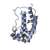 4o71C  4o74C 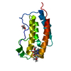 4o75C 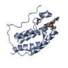 4o76C 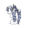 4o77C 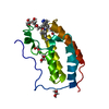 4o78C 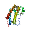 4o7aC 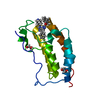 4o7bC 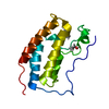 4o7cC 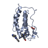 4o7eC 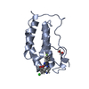 4o7fC 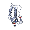 4ps5C 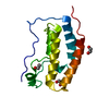 2ossS C: citing same article ( S: Starting model for refinement |
|---|---|
| Similar structure data |


-Links
 PDBj
PDBj- Assembly
| Deposited unit | 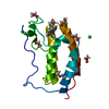
| ||||||||
|---|---|---|---|---|---|---|---|---|---|
| 1 |
| ||||||||
| Unit cell |
|
-Components
-Protein , 1 types, 1 molecules A
| #1: Protein | Mass: 15099.380 Da / Num. of mol.: 1 / Fragment: UNP residues 44-168 Source method: isolated from a genetically manipulated source Source: (gene. exp.) Homo sapiens (human) / Gene: BRD4, HUNK1 / Plasmid: PNIC28-BSA4 / Production host:  |
|---|
-Non-polymers , 5 types, 203 molecules 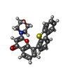


| #2: Chemical | ChemComp-2R4 /  Mass: 413.488 Da / Num. of mol.: 1 / Source method: obtained synthetically / Formula: C25H19NO3S Mass: 413.488 Da / Num. of mol.: 1 / Source method: obtained synthetically / Formula: C25H19NO3S | ||||
|---|---|---|---|---|---|
| #3: Chemical | ChemComp-PEG /  Mass: 106.120 Da / Num. of mol.: 1 / Source method: obtained synthetically / Formula: C4H10O3 Mass: 106.120 Da / Num. of mol.: 1 / Source method: obtained synthetically / Formula: C4H10O3 | ||||
| #4: Chemical | ChemComp-EDO /  Mass: 62.068 Da / Num. of mol.: 5 / Source method: obtained synthetically / Formula: C2H6O2 Mass: 62.068 Da / Num. of mol.: 5 / Source method: obtained synthetically / Formula: C2H6O2#5: Chemical |  Mass: 35.453 Da / Num. of mol.: 3 / Source method: obtained synthetically / Formula: Cl Mass: 35.453 Da / Num. of mol.: 3 / Source method: obtained synthetically / Formula: Cl#6: Water | ChemComp-HOH / | Mass: 18.015 Da / Num. of mol.: 193 / Source method: isolated from a natural source / Formula: H2O |
-Experimental details
-Experiment
| Experiment | Method: X-RAY DIFFRACTION / Number of used crystals: 1 |
|---|
- Sample preparation
| Crystal | Density Matthews: 2.14 Å3/Da / Density % sol: 42.59 % |
|---|---|
| Crystal grow | Temperature: 291 K / Method: vapor diffusion, hanging drop / pH: 7.5 Details: 12.5 MG/ML BRD4, 5MM HEPES pH 7.5, 50MM SODIUM CHLORIDE, 0.5MM DTT, 50MM TRIS PH8.5, 0.1M AMMONIUM SULFATE, 12.5% PEG 3,350, 10% DMSO, 1 MM NU7441, VAPOR DIFFUSION, HANGING DROP, temperature 291K |
-Data collection
| Diffraction | Mean temperature: 93 K |
|---|---|
| Diffraction source | Source: ROTATING ANODE / Type: RIGAKU MICROMAX-007 HF / Wavelength: 1.54178 Å |
| Detector | Type: RIGAKU SATURN 944+ / Detector: CCD / Date: Nov 22, 2013 / Details: MIRRORS |
| Radiation | Monochromator: MIRRORS / Protocol: SINGLE WAVELENGTH / Monochromatic (M) / Laue (L): M / Scattering type: x-ray |
| Radiation wavelength | Wavelength: 1.54178 Å / Relative weight: 1 |
| Reflection | Resolution: 1.4→20 Å / Num. obs: 26039 / % possible obs: 99 % / Observed criterion σ(I): -3 / Redundancy: 3.4 % / Rsym value: 0.03 / Net I/σ(I): 25.4 |
| Reflection shell | Resolution: 1.4→1.44 Å / Redundancy: 1.8 % / Mean I/σ(I) obs: 4 / Rsym value: 0.166 / % possible all: 92.6 |
- Processing
| Software |
| ||||||||||||||||||||||||||||||||||||||||||||||||||||||||||||||||||||||
|---|---|---|---|---|---|---|---|---|---|---|---|---|---|---|---|---|---|---|---|---|---|---|---|---|---|---|---|---|---|---|---|---|---|---|---|---|---|---|---|---|---|---|---|---|---|---|---|---|---|---|---|---|---|---|---|---|---|---|---|---|---|---|---|---|---|---|---|---|---|---|---|
| Refinement | Method to determine structure: MOLECULAR REPLACEMENT Starting model: PDB entry 2OSS Resolution: 1.4→19.652 Å / SU ML: 0.11 / σ(F): 1.37 / Phase error: 13.79 / Stereochemistry target values: ML
| ||||||||||||||||||||||||||||||||||||||||||||||||||||||||||||||||||||||
| Solvent computation | Shrinkage radii: 0.9 Å / VDW probe radii: 1.11 Å / Solvent model: FLAT BULK SOLVENT MODEL | ||||||||||||||||||||||||||||||||||||||||||||||||||||||||||||||||||||||
| Refinement step | Cycle: LAST / Resolution: 1.4→19.652 Å
| ||||||||||||||||||||||||||||||||||||||||||||||||||||||||||||||||||||||
| Refine LS restraints |
| ||||||||||||||||||||||||||||||||||||||||||||||||||||||||||||||||||||||
| LS refinement shell |
|
