Mass: 18.015 Da / Num. of mol.: 123 / Source method: isolated from a natural source / Formula: H2O
Has protein modification
Y
Nonpolymer details
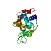
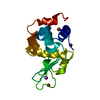
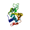
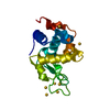
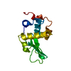
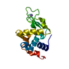
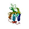
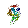
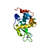
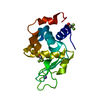
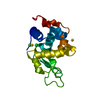
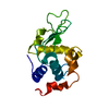
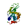
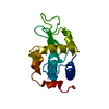
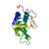
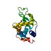
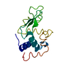
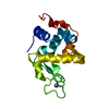
THE X-RAY STRUCTURE REVEALS THAT NAMI-A BINDS THE PROTEIN, AS NAKED RUTHENIUM ION, AT TWO DISTINCT ...THE X-RAY STRUCTURE REVEALS THAT NAMI-A BINDS THE PROTEIN, AS NAKED RUTHENIUM ION, AT TWO DISTINCT SITES (EITHER TO ASP101 OR TO ASP119), AFTER RELEASING ALL ITS ORIGINAL LIGANDS (DMSO, IMIDAZOLE AND CL-). THIS INDICATES THAT NAMI-A UNDERGOES DRAMATIC CHANGES IN THE COORDINATION ENVIRONMENT OF THE RUTHENIUM CENTRE UPON THE PROTEIN BINDING AND THAT ASP SIDE CHAINS ARE PREFERENTIAL TARGET SITES FOR RUTHENIUM BINDING TO THE PROTEIN
-
Experimental details
-
Experiment
Experiment
Method: X-RAY DIFFRACTION / Number of used crystals: 1
-
Sample preparation
Crystal
Density Matthews: 1.97 Å3/Da / Density % sol: 37.52 %
Crystal grow
Temperature: 293 K / Method: vapor diffusion, hanging drop / pH: 4.5 Details: Crystals of the HEWL/NAMI-A adduct suitable for X-ray diffraction studies Have been grown by hanging drop vapor diffusion by mixing 1 L of 20 mg/mL HEWL/NAMI-A complex (protein to metal ...Details: Crystals of the HEWL/NAMI-A adduct suitable for X-ray diffraction studies Have been grown by hanging drop vapor diffusion by mixing 1 L of 20 mg/mL HEWL/NAMI-A complex (protein to metal ratio 1:10) with an equal volume of reservoir solution containing 1.0 M NaCl and 50 mM sodium acetate pH 4.5. These crystals were fished with nylon loops and flash-frozen at 100 K using nitrogen gas, without cryoprotectant. This procedure partly dehydrates the crystals, enhancing in some cases the resolution of X-ray diffraction data, VAPOR DIFFUSION, HANGING DROP, temperature 293K
Resolution: 1.85→22.15 Å / Cor.coef. Fo:Fc: 0.955 / Cor.coef. Fo:Fc free: 0.936 / SU B: 3.321 / SU ML: 0.102 / Cross valid method: THROUGHOUT / ESU R: 0.163 / ESU R Free: 0.159 / Stereochemistry target values: MAXIMUM LIKELIHOOD / Details: HYDROGENS HAVE BEEN ADDED IN THE RIDING POSITIONS
Rfactor
Num. reflection
% reflection
Selection details
Rfree
0.23994
490
4.8 %
RANDOM
Rwork
0.17626
-
-
-
obs
0.17933
9628
98.91 %
-
Solvent computation
Ion probe radii: 0.8 Å / Shrinkage radii: 0.8 Å / VDW probe radii: 1.2 Å / Solvent model: MASK
 Movie
Movie Controller

 Yorodumi
 Open data
 Basic information
 Components
 Keywords
 Function and homology information
 X-RAY DIFFRACTION /
  Authors
 Citation
 Structure visualization
 Downloads & links
 Other downloads


 PDBj
PDBj


 Assembly
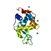

 Mass: 101.070 Da / Num. of mol.: 2 / Source method: obtained synthetically / Formula: Ru
 Mass: 35.453 Da / Num. of mol.: 2 / Source method: obtained synthetically / Formula: Cl
 Mass: 22.990 Da / Num. of mol.: 1 / Source method: obtained synthetically / Formula: Na
 Mass: 18.015 Da / Num. of mol.: 123 / Source method: isolated from a natural source / Formula: H2O
 Sample preparation
 Processing