[English] 日本語
Yorodumi
- PDB-4noo: Molecular mechanism for self-protection against type VI secretion... -

+
Open data


ID or keywords:

Loading...

-
Basic information

Entry
Database: PDB / ID: 4noo
Title
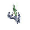
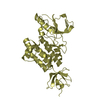
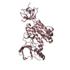
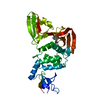
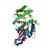
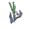
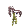
Molecular mechanism for self-protection against type VI secretion system in Vibrio cholerae
Components
  • Putative uncharacterized protein
  • VgrG protein
KeywordsIMMUNE SYSTEM / self-protection / chitosanase-fold / three-helical bundle / glycoside hydrolase effector / immunity protein
Function / homology
Function and homology information


Hydrolases; Glycosylases; Glycosidases, i.e. enzymes that hydrolyse O- and S-glycosyl compounds / hydrolase activity / extracellular region / identical protein binding
Similarity search - Function
: / Type VI secretion system spike protein VgrG3-like, C-terminal / Helicase, Ruva Protein; domain 3 - #1160 / : / : / Antitoxin TsiV3 / Type VI secretion system, RhsGE-associated Vgr family subset / Phage tail baseplate hub (GPD) / Type VI secretion system, RhsGE-associated Vgr protein / Gp5/Type VI secretion system Vgr protein, OB-fold domain ...: / Type VI secretion system spike protein VgrG3-like, C-terminal / Helicase, Ruva Protein; domain 3 - #1160 / : / : / Antitoxin TsiV3 / Type VI secretion system, RhsGE-associated Vgr family subset / Phage tail baseplate hub (GPD) / Type VI secretion system, RhsGE-associated Vgr protein / Gp5/Type VI secretion system Vgr protein, OB-fold domain / Type VI secretion system/phage-baseplate injector OB domain / Vgr protein, OB-fold domain superfamily / PGBD superfamily / Peptidoglycan binding-like / Putative peptidoglycan binding domain / PGBD-like superfamily / Helicase, Ruva Protein; domain 3 / Orthogonal Bundle / Mainly Alpha
Similarity search - Domain/homology
Antitoxin protein TsiV3 / Type VI secretion system spike protein VgrG3
Similarity search - Component
Biological speciesVibrio cholerae O1 biovar El Tor (bacteria)
MethodX-RAY DIFFRACTION / SYNCHROTRON / SAD / Resolution: 2.3 Å
AuthorsYang, X. / Xu, M. / Jiang, T. / Fan, Z.
CitationJournal: Acta Crystallogr.,Sect.D / Year: 2014
Title: Molecular mechanism for self-protection against the type VI secretion system in Vibrio cholerae.
Authors: Yang, X. / Xu, M. / Wang, Y. / Xia, P. / Wang, S. / Ye, B. / Tong, L. / Jiang, T. / Fan, Z.
History
DepositionNov 20, 2013Deposition site: RCSB / Processing site: RCSB
Revision 1.0Apr 9, 2014Provider: repository / Type: Initial release
Revision 1.1Apr 16, 2014Group: Database references

-
Structure visualization

Structure viewerMolecule:
MolmilJmol/JSmol

Downloads & links

-
Assembly

Deposited unit
A: VgrG protein
B: Putative uncharacterized protein
C: VgrG protein
D: Putative uncharacterized protein


Theoretical massNumber of molelcules
Total (without water)69,8694
Polymers69,8694
Non-polymers00
Water9,458525
1
A: VgrG protein
B: Putative uncharacterized protein


Theoretical massNumber of molelcules
Total (without water)34,9342
Polymers34,9342
Non-polymers00
Water362
TypeNameSymmetry operationNumber
identity operation1_555x,y,z1
Buried area2510 Å2
ΔGint-12 kcal/mol
Surface area14960 Å2
MethodPISA
2
C: VgrG protein
D: Putative uncharacterized protein


Theoretical massNumber of molelcules
Total (without water)34,9342
Polymers34,9342
Non-polymers00
Water362
TypeNameSymmetry operationNumber
identity operation1_555x,y,z1
Buried area2550 Å2
ΔGint-12 kcal/mol
Surface area14970 Å2
MethodPISA
Unit cell
Length a, b, c (Å)52.502, 116.034, 68.225
Angle α, β, γ (deg.)90.00, 94.05, 90.00
Int Tables number4
Space group name H-MP1211

-
Components

#1: Protein VgrG protein


Mass: 23576.129 Da / Num. of mol.: 2 / Fragment: VgrG3C catalytic domain
Source method: isolated from a genetically manipulated source
Source: (gene. exp.) Vibrio cholerae O1 biovar El Tor (bacteria)
Strain: ATCC 39315 / El Tor Inaba N16961 / Gene: VC_A0123 / Plasmid: pRSFduet / Production host: Escherichia coli (E. coli) / Strain (production host): BL21-CodonPlus (DE3)-RIPL / References: UniProt: Q9KN42
#2: Protein Putative uncharacterized protein


Mass: 11358.249 Da / Num. of mol.: 2
Fragment: TsiV3 without the predicted 24aa N-terminal periplasm localization signal
Source method: isolated from a genetically manipulated source
Source: (gene. exp.) Vibrio cholerae O1 biovar El Tor (bacteria)
Strain: ATCC 39315 / El Tor Inaba N16961 / Gene: VC_A0124 / Plasmid: pRSFduet / Production host: Escherichia coli (E. coli) / Strain (production host): BL21-CodonPlus (DE3)-RIPL / References: UniProt: Q9KN41
#3: Water ChemComp-HOH / water


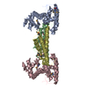
Mass: 18.015 Da / Num. of mol.: 525 / Source method: isolated from a natural source / Formula: H2O

-
Experimental details

-
Experiment

ExperimentMethod: X-RAY DIFFRACTION / Number of used crystals: 1

-
Sample preparation

CrystalDensity Matthews: 2.97 Å3/Da / Density % sol: 58.54 %
Crystal growTemperature: 289 K / Method: vapor diffusion, hanging drop / pH: 5.6
Details: 15% (m/v) PEG8000, 0.1M Sodium Cacodylate pH5.6, 0.2M Mg acetate, VAPOR DIFFUSION, HANGING DROP, temperature 289K

-
Data collection

DiffractionMean temperature: 100 K
Diffraction sourceSource: SYNCHROTRON / Site: BSRF / Beamline: 3W1A / Wavelength: 0.9789 Å
DetectorType: MAR CCD 165 mm / Detector: CCD / Date: May 11, 2013
RadiationMonochromator: GRAPHITE / Protocol: SINGLE WAVELENGTH / Monochromatic (M) / Laue (L): M / Scattering type: x-ray
Radiation wavelengthWavelength: 0.9789 Å / Relative weight: 1
ReflectionResolution: 2.3→29.5 Å / Num. all: 34369 / Num. obs: 34369 / % possible obs: 99.92 % / Observed criterion σ(F): 0 / Observed criterion σ(I): 0 / Redundancy: 7.7 % / Rmerge(I) obs: 0.072 / Net I/σ(I): 28
Reflection shellResolution: 2.3→2.34 Å / Redundancy: 7.7 % / Rmerge(I) obs: 0.422 / Mean I/σ(I) obs: 4.9 / % possible all: 100

-
Processing

Software
NameVersionClassification
ADSCQuantumdata collection
PHENIXmodel building
REFMAC5.7.0029refinement
HKL-2000data reduction
HKL-2000data scaling
PHENIXphasing
RefinementMethod to determine structure: SAD / Resolution: 2.3→29.5 Å / Cor.coef. Fo:Fc: 0.946 / Cor.coef. Fo:Fc free: 0.913 / Cross valid method: THROUGHOUT / ESU R: 0.232 / ESU R Free: 0.215 / Stereochemistry target values: MAXIMUM LIKELIHOOD / Details: HYDROGENS HAVE BEEN ADDED IN THE RIDING POSITIONS
RfactorNum. reflection% reflectionSelection details
Rfree0.23441 1822 5 %RANDOM
Rwork0.18629 ---
obs0.18868 34369 99.92 %-
all-34369 --
Solvent computationIon probe radii: 0.8 Å / Shrinkage radii: 0.8 Å / VDW probe radii: 1.2 Å / Solvent model: MASK
Displacement parametersBiso mean: 20.487 Å2
Baniso -1Baniso -2Baniso -3
1-0.57 Å20 Å2-0.05 Å2
2---1.13 Å20 Å2
3---0.56 Å2
Refinement stepCycle: LAST / Resolution: 2.3→29.5 Å
ProteinNucleic acidLigandSolventTotal
Num. atoms4734 0 0 525 5259
Refine LS restraints
Refine-IDTypeDev idealDev ideal targetNumber
X-RAY DIFFRACTIONr_bond_refined_d0.010.0194830
X-RAY DIFFRACTIONr_bond_other_d00.024546
X-RAY DIFFRACTIONr_angle_refined_deg1.2781.9516508
X-RAY DIFFRACTIONr_angle_other_deg3.5973.00210454
X-RAY DIFFRACTIONr_dihedral_angle_1_deg5.3895586
X-RAY DIFFRACTIONr_dihedral_angle_2_deg32.74725.041246
X-RAY DIFFRACTIONr_dihedral_angle_3_deg13.6715842
X-RAY DIFFRACTIONr_dihedral_angle_4_deg18.6321524
X-RAY DIFFRACTIONr_chiral_restr0.0740.2690
X-RAY DIFFRACTIONr_gen_planes_refined0.0050.025542
X-RAY DIFFRACTIONr_gen_planes_other0.0080.021158
X-RAY DIFFRACTIONr_nbd_refined
X-RAY DIFFRACTIONr_nbd_other
X-RAY DIFFRACTIONr_nbtor_refined
X-RAY DIFFRACTIONr_nbtor_other
X-RAY DIFFRACTIONr_xyhbond_nbd_refined
X-RAY DIFFRACTIONr_xyhbond_nbd_other
X-RAY DIFFRACTIONr_metal_ion_refined
X-RAY DIFFRACTIONr_metal_ion_other
X-RAY DIFFRACTIONr_symmetry_vdw_refined
X-RAY DIFFRACTIONr_symmetry_vdw_other
X-RAY DIFFRACTIONr_symmetry_hbond_refined
X-RAY DIFFRACTIONr_symmetry_hbond_other
X-RAY DIFFRACTIONr_symmetry_metal_ion_refined
X-RAY DIFFRACTIONr_symmetry_metal_ion_other
X-RAY DIFFRACTIONr_mcbond_it
X-RAY DIFFRACTIONr_mcbond_other
X-RAY DIFFRACTIONr_mcangle_it
X-RAY DIFFRACTIONr_mcangle_other
X-RAY DIFFRACTIONr_scbond_it
X-RAY DIFFRACTIONr_scbond_other
X-RAY DIFFRACTIONr_scangle_it
X-RAY DIFFRACTIONr_scangle_other
X-RAY DIFFRACTIONr_long_range_B_refined
X-RAY DIFFRACTIONr_long_range_B_other
X-RAY DIFFRACTIONr_rigid_bond_restr
X-RAY DIFFRACTIONr_sphericity_free
X-RAY DIFFRACTIONr_sphericity_bonded
LS refinement shellResolution: 2.3→2.359 Å / Total num. of bins used: 20
RfactorNum. reflection% reflection
Rfree0.344 115 -
Rwork0.233 2539 -
obs--99.66 %

+
About Yorodumi

-
News

-
Feb 9, 2022. New format data for meta-information of EMDB entries

New format data for meta-information of EMDB entries

  • Version 3 of the EMDB header file is now the official format.
  • The previous official version 1.9 will be removed from the archive.

Related info.:EMDB header

External links:wwPDB to switch to version 3 of the EMDB data model

-
Aug 12, 2020. Covid-19 info

Covid-19 info

URL: https://pdbjlvh1.pdbj.org/emnavi/covid19.php

New page: Covid-19 featured information page in EM Navigator.

Related info.:Covid-19 info / Mar 5, 2020. Novel coronavirus structure data

+
Mar 5, 2020. Novel coronavirus structure data

Novel coronavirus structure data

Related info.:Yorodumi Speices / Aug 12, 2020. Covid-19 info

External links:COVID-19 featured content - PDBj / Molecule of the Month (242):Coronavirus Proteases

+
Jan 31, 2019. EMDB accession codes are about to change! (news from PDBe EMDB page)

EMDB accession codes are about to change! (news from PDBe EMDB page)

  • The allocation of 4 digits for EMDB accession codes will soon come to an end. Whilst these codes will remain in use, new EMDB accession codes will include an additional digit and will expand incrementally as the available range of codes is exhausted. The current 4-digit format prefixed with “EMD-” (i.e. EMD-XXXX) will advance to a 5-digit format (i.e. EMD-XXXXX), and so on. It is currently estimated that the 4-digit codes will be depleted around Spring 2019, at which point the 5-digit format will come into force.
  • The EM Navigator/Yorodumi systems omit the EMD- prefix.

Related info.:Q: What is EMD? / ID/Accession-code notation in Yorodumi/EM Navigator

External links:EMDB Accession Codes are Changing Soon! / Contact to PDBj

+
Jul 12, 2017. Major update of PDB

Major update of PDB

  • wwPDB released updated PDB data conforming to the new PDBx/mmCIF dictionary.
  • This is a major update changing the version number from 4 to 5, and with Remediation, in which all the entries are updated.
  • In this update, many items about electron microscopy experimental information are reorganized (e.g. em_software).
  • Now, EM Navigator and Yorodumi are based on the updated data.

External links:wwPDB Remediation / Enriched Model Files Conforming to OneDep Data Standards Now Available in the PDB FTP Archive

-
Yorodumi

Thousand views of thousand structures

  • Yorodumi is a browser for structure data from EMDB, PDB, SASBDB, etc.
  • This page is also the successor to EM Navigator detail page, and also detail information page/front-end page for Omokage search.
  • The word "yorodu" (or yorozu) is an old Japanese word meaning "ten thousand". "mi" (miru) is to see.

Related info.:EMDB / PDB / SASBDB / Comparison of 3 databanks / Yorodumi Search / Aug 31, 2016. New EM Navigator & Yorodumi / Yorodumi Papers / Jmol/JSmol / Function and homology information / Changes in new EM Navigator and Yorodumi

Read more