[English] 日本語
Yorodumi
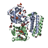
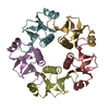
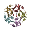
- PDB-4nl3: Crystal Structure of Listeria monocytogenes Hfq in complex with U6 RNA -

+
Open data


ID or keywords:

Loading...

-
Basic information

Entry
Database: PDB / ID: 4nl3
TitleCrystal Structure of Listeria monocytogenes Hfq in complex with U6 RNA
Components
  • 5'-R(*UP*UP*UP*UP*UP*U)-3'
  • Protein hfq
KeywordsRNA BINDING PROTEIN/RNA / LSm/Sm proteins / RNA chaperone / sRNA / RNA BINDING PROTEIN-RNA complex
Function / homology
Function and homology information


regulation of translation, ncRNA-mediated / regulation of RNA stability / regulation of DNA-templated transcription / RNA binding / cytosol
Similarity search - Function
RNA-binding protein Hfq / Hfq protein / SH3 type barrels. - #100 / : / Sm domain profile. / LSM domain superfamily / SH3 type barrels. / Roll / Mainly Beta
Similarity search - Domain/homology
RNA / : / RNA-binding protein Hfq
Similarity search - Component
Biological speciesListeria monocytogenes (bacteria)
MethodX-RAY DIFFRACTION / SYNCHROTRON / MOLECULAR REPLACEMENT / Resolution: 3.1 Å
AuthorsKovach, A.R. / Brennan, R.G.
CitationJournal: Rna / Year: 2014
Title: Recognition of U-rich RNA by Hfq from the Gram-positive pathogen Listeria monocytogenes.
Authors: Kovach, A.R. / Hoff, K.E. / Canty, J.T. / Orans, J. / Brennan, R.G.
History
DepositionNov 13, 2013Deposition site: RCSB / Processing site: RCSB
Revision 1.0Sep 10, 2014Provider: repository / Type: Initial release
Revision 1.1Oct 1, 2014Group: Database references
Revision 1.2Nov 22, 2017Group: Refinement description / Structure summary / Category: audit_author / software
Item: _audit_author.name / _software.classification ..._audit_author.name / _software.classification / _software.contact_author / _software.contact_author_email / _software.date / _software.language / _software.location / _software.name / _software.type / _software.version
Revision 1.3Sep 20, 2023Group: Data collection / Database references / Refinement description
Category: chem_comp_atom / chem_comp_bond ...chem_comp_atom / chem_comp_bond / database_2 / pdbx_initial_refinement_model / struct_ncs_dom_lim
Item: _database_2.pdbx_DOI / _database_2.pdbx_database_accession ..._database_2.pdbx_DOI / _database_2.pdbx_database_accession / _struct_ncs_dom_lim.beg_auth_comp_id / _struct_ncs_dom_lim.beg_label_asym_id / _struct_ncs_dom_lim.beg_label_comp_id / _struct_ncs_dom_lim.beg_label_seq_id / _struct_ncs_dom_lim.end_auth_comp_id / _struct_ncs_dom_lim.end_label_asym_id / _struct_ncs_dom_lim.end_label_comp_id / _struct_ncs_dom_lim.end_label_seq_id

-
Structure visualization

Structure viewerMolecule:
MolmilJmol/JSmol

Downloads & links

-
Assembly

Deposited unit
D: Protein hfq
A: Protein hfq
B: Protein hfq
C: Protein hfq
E: Protein hfq
F: Protein hfq
J: Protein hfq
G: Protein hfq
H: Protein hfq
I: Protein hfq
K: Protein hfq
L: Protein hfq
R: 5'-R(*UP*UP*UP*UP*UP*U)-3'
Z: 5'-R(*UP*UP*UP*UP*UP*U)-3'


Theoretical massNumber of molelcules
Total (without water)108,28414
Polymers108,28414
Non-polymers00
Water362
1
D: Protein hfq
A: Protein hfq
B: Protein hfq
J: Protein hfq
G: Protein hfq
H: Protein hfq
R: 5'-R(*UP*UP*UP*UP*UP*U)-3'


Theoretical massNumber of molelcules
Total (without water)54,1427
Polymers54,1427
Non-polymers00
Water362
TypeNameSymmetry operationNumber
identity operation1_555x,y,z1
Buried area13740 Å2
ΔGint-119 kcal/mol
Surface area19100 Å2
MethodPISA
2
C: Protein hfq
E: Protein hfq
F: Protein hfq
I: Protein hfq
K: Protein hfq
L: Protein hfq
Z: 5'-R(*UP*UP*UP*UP*UP*U)-3'


Theoretical massNumber of molelcules
Total (without water)54,1427
Polymers54,1427
Non-polymers00
Water0
TypeNameSymmetry operationNumber
identity operation1_555x,y,z1
Buried area13320 Å2
ΔGint-89 kcal/mol
Surface area20720 Å2
MethodPISA
Unit cell
Length a, b, c (Å)124.028, 123.934, 67.595
Angle α, β, γ (deg.)90.000, 90.060, 90.000
Int Tables number5
Space group name H-MC121
Noncrystallographic symmetry (NCS)NCS domain:
IDEns-IDDetails
11chain A
21chain B
31chain C
41chain D
51chain E
61chain F
71chain G
81chain H
91chain I
101chain J
111chain K
121chain L
12chain R
22chain Z

NCS domain segments:

Component-ID: 1

Dom-IDEns-IDBeg auth comp-IDBeg label comp-IDEnd auth comp-IDEnd label comp-IDSelection detailsAuth asym-IDLabel asym-IDAuth seq-IDLabel seq-ID
11LYSLYSASNASNchain AAB2 - 732 - 73
21LYSLYSLEULEUchain BBC2 - 722 - 72
31METMETPROPROchain CCD1 - 741 - 74
41METMETASNASNchain DDA1 - 731 - 73
51LYSLYSASNASNchain EEE2 - 732 - 73
61METMETASPASPchain FFF1 - 751 - 75
71METMETASNASNchain GGH1 - 731 - 73
81LYSLYSLEULEUchain HHI2 - 722 - 72
91METMETPROPROchain IIJ1 - 741 - 74
101METMETASNASNchain JJG1 - 731 - 73
111METMETASNASNchain KKK1 - 731 - 73
121METMETPROPROchain LLL1 - 741 - 74
12UUUUchain RRM17 - 221 - 6
22UUUUchain ZZN27 - 321 - 6

NCS ensembles :
ID
1
2

-
Components

#1: Protein
Protein hfq


Mass: 8724.953 Da / Num. of mol.: 12
Source method: isolated from a genetically manipulated source
Source: (gene. exp.) Listeria monocytogenes (bacteria) / Gene: hfq, LMHCC_1277 / Production host: Escherichia coli (E. coli) / References: UniProt: B8DG33, UniProt: Q92C58*PLUS
#2: RNA chain 5'-R(*UP*UP*UP*UP*UP*U)-3'


Mass: 1792.037 Da / Num. of mol.: 2 / Source method: obtained synthetically
#3: Water ChemComp-HOH / water


Mass: 18.015 Da / Num. of mol.: 2 / Source method: isolated from a natural source / Formula: H2O

-
Experimental details

-
Experiment

ExperimentMethod: X-RAY DIFFRACTION / Number of used crystals: 1

-
Sample preparation

CrystalDensity Matthews: 2.4 Å3/Da / Density % sol: 48.72 %
Crystal growTemperature: 298 K / Method: vapor diffusion, hanging drop / pH: 7.5
Details: 40% 1,2-propanediol, 100 mM HEPES, pH 7.5, VAPOR DIFFUSION, HANGING DROP, temperature 298K

-
Data collection

DiffractionMean temperature: 100 K
Diffraction sourceSource: SYNCHROTRON / Site: APS / Beamline: 22-ID / Wavelength: 1 Å
DetectorType: MARMOSAIC 300 mm CCD / Detector: CCD / Date: Jul 29, 2013
RadiationMonochromator: Rosenbaum-Rock double-crystal Si(111) / Protocol: SINGLE WAVELENGTH / Monochromatic (M) / Laue (L): M / Scattering type: x-ray
Radiation wavelengthWavelength: 1 Å / Relative weight: 1
ReflectionResolution: 3.1→50 Å / Num. obs: 17828 / % possible obs: 95.9 % / Redundancy: 3.3 % / Biso Wilson estimate: 53.28 Å2 / Rmerge(I) obs: 0.114 / Χ2: 1.666 / Net I/σ(I): 7.4
Reflection shell
Resolution (Å)Redundancy (%)Rmerge(I) obsNum. unique allΧ2Diffraction-ID% possible all
3.1-3.152.10.3036391.114170.5
3.15-3.212.30.2637491.346179
3.21-3.272.50.2697961.049187
3.27-3.342.70.2478631.258192.6
3.34-3.4130.2298551.373195.5
3.41-3.493.20.2048951.328197
3.49-3.583.30.2119441.549199.4
3.58-3.683.40.2099241.666199.5
3.68-3.783.60.179031.643199.9
3.78-3.913.60.1839461.4561100
3.91-4.043.70.1529031.526199.8
4.04-4.213.70.1149341.6131100
4.21-4.43.60.0959451.9621100
4.4-4.633.60.0869221.908199.9
4.63-4.923.60.0949202.038199.9
4.92-5.33.60.0989421.831199.8
5.3-5.833.60.1139451.888199.8
5.83-6.673.60.0989132.011199.8
6.67-8.43.50.0739461.794199.8
8.4-503.30.0419441.791198.1

-
Processing

Software
NameVersionClassificationNB
DENZOdata reduction
SCALEPACKdata scaling
PHENIX1.8.2_1309refinement
PDB_EXTRACT3.11data extraction
SERGUIdata collection
HKL-2000data reduction
PHASERphasing
RefinementMethod to determine structure: MOLECULAR REPLACEMENT
Starting model: PDB ENTRY 4NL2
Resolution: 3.1→45.675 Å / FOM work R set: 0.7677 / SU ML: 0.38 / σ(F): 1.34 / Phase error: 29.81 / Stereochemistry target values: ML
RfactorNum. reflection% reflection
Rfree0.2827 907 5.11 %
Rwork0.2211 16853 -
obs0.2242 17760 95.47 %
Solvent computationShrinkage radii: 0.9 Å / VDW probe radii: 1.11 Å / Solvent model: FLAT BULK SOLVENT MODEL
Displacement parametersBiso max: 113.01 Å2 / Biso mean: 36.02 Å2 / Biso min: 11.82 Å2
Refinement stepCycle: LAST / Resolution: 3.1→45.675 Å
ProteinNucleic acidLigandSolventTotal
Num. atoms6932 234 0 2 7168
Refine LS restraints
Refine-IDTypeDev idealNumber
X-RAY DIFFRACTIONf_bond_d0.017292
X-RAY DIFFRACTIONf_angle_d1.1829859
X-RAY DIFFRACTIONf_chiral_restr0.0691137
X-RAY DIFFRACTIONf_plane_restr0.0051236
X-RAY DIFFRACTIONf_dihedral_angle_d16.6062733
Refine LS restraints NCS
Ens-IDDom-IDAuth asym-IDNumberRefine-IDRmsType
11A4242X-RAY DIFFRACTION20.533TORSIONAL
12B4242X-RAY DIFFRACTION20.533TORSIONAL
13C4242X-RAY DIFFRACTION20.533TORSIONAL
14D4242X-RAY DIFFRACTION20.533TORSIONAL
15E4242X-RAY DIFFRACTION20.533TORSIONAL
16F4242X-RAY DIFFRACTION20.533TORSIONAL
17G4242X-RAY DIFFRACTION20.533TORSIONAL
18H4242X-RAY DIFFRACTION20.533TORSIONAL
19I4242X-RAY DIFFRACTION20.533TORSIONAL
110J4242X-RAY DIFFRACTION20.533TORSIONAL
111K4242X-RAY DIFFRACTION20.533TORSIONAL
112L4242X-RAY DIFFRACTION20.533TORSIONAL
21R136X-RAY DIFFRACTION20.533TORSIONAL
22Z136X-RAY DIFFRACTION20.533TORSIONAL
LS refinement shell

Refine-ID: X-RAY DIFFRACTION / Total num. of bins used: 6

Resolution (Å)Rfactor RfreeNum. reflection RfreeRfactor RworkNum. reflection RworkNum. reflection all% reflection obs (%)
3.0997-3.29380.3711240.25272319244380
3.2938-3.54810.34411570.24842838299596
3.5481-3.9050.30461500.232229103060100
3.905-4.46960.2541520.204529453097100
4.4696-5.62960.25931610.202129273088100
5.6296-45.67950.25481630.22232914307798

+
About Yorodumi

-
News

-
Feb 9, 2022. New format data for meta-information of EMDB entries

New format data for meta-information of EMDB entries

  • Version 3 of the EMDB header file is now the official format.
  • The previous official version 1.9 will be removed from the archive.

Related info.:EMDB header

External links:wwPDB to switch to version 3 of the EMDB data model

-
Aug 12, 2020. Covid-19 info

Covid-19 info

URL: https://pdbj.org/emnavi/covid19.php

New page: Covid-19 featured information page in EM Navigator.

Related info.:Covid-19 info / Mar 5, 2020. Novel coronavirus structure data

+
Mar 5, 2020. Novel coronavirus structure data

Novel coronavirus structure data

Related info.:Yorodumi Speices / Aug 12, 2020. Covid-19 info

External links:COVID-19 featured content - PDBj / Molecule of the Month (242):Coronavirus Proteases

+
Jan 31, 2019. EMDB accession codes are about to change! (news from PDBe EMDB page)

EMDB accession codes are about to change! (news from PDBe EMDB page)

  • The allocation of 4 digits for EMDB accession codes will soon come to an end. Whilst these codes will remain in use, new EMDB accession codes will include an additional digit and will expand incrementally as the available range of codes is exhausted. The current 4-digit format prefixed with “EMD-” (i.e. EMD-XXXX) will advance to a 5-digit format (i.e. EMD-XXXXX), and so on. It is currently estimated that the 4-digit codes will be depleted around Spring 2019, at which point the 5-digit format will come into force.
  • The EM Navigator/Yorodumi systems omit the EMD- prefix.

Related info.:Q: What is EMD? / ID/Accession-code notation in Yorodumi/EM Navigator

External links:EMDB Accession Codes are Changing Soon! / Contact to PDBj

+
Jul 12, 2017. Major update of PDB

Major update of PDB

  • wwPDB released updated PDB data conforming to the new PDBx/mmCIF dictionary.
  • This is a major update changing the version number from 4 to 5, and with Remediation, in which all the entries are updated.
  • In this update, many items about electron microscopy experimental information are reorganized (e.g. em_software).
  • Now, EM Navigator and Yorodumi are based on the updated data.

External links:wwPDB Remediation / Enriched Model Files Conforming to OneDep Data Standards Now Available in the PDB FTP Archive

-
Yorodumi

Thousand views of thousand structures

  • Yorodumi is a browser for structure data from EMDB, PDB, SASBDB, etc.
  • This page is also the successor to EM Navigator detail page, and also detail information page/front-end page for Omokage search.
  • The word "yorodu" (or yorozu) is an old Japanese word meaning "ten thousand". "mi" (miru) is to see.

Related info.:EMDB / PDB / SASBDB / Comparison of 3 databanks / Yorodumi Search / Aug 31, 2016. New EM Navigator & Yorodumi / Yorodumi Papers / Jmol/JSmol / Function and homology information / Changes in new EM Navigator and Yorodumi

Read more