 Movie
Movie Controller

[English] 日本語
 Yorodumi
- PDB-4nhw: Crystal structure of glutathione transferase SMc00097 from Sinorh... -
+ Open data

- Basic information
| Entry | Database: PDB / ID: 4nhw | ||||||
|---|---|---|---|---|---|---|---|
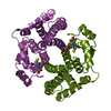
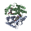
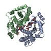
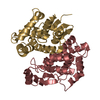
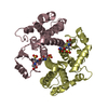
| Title | Crystal structure of glutathione transferase SMc00097 from Sinorhizobium meliloti, target EFI-507275, with one glutathione bound per one protein subunit | ||||||
 Components Components | Glutathione S-transferase | ||||||
 Keywords Keywords | TRANSFERASE / glutathione S-transferase / Enzyme Function Initiative / EFI / structural genomics | ||||||
| Function / homology |  Function and homology information Function and homology informationGlutathione S-transferase Yfyf (Class Pi); Chain A, domain 2 - #10 / Glutathione S-transferase Yfyf (Class Pi); Chain A, domain 2 / Glutaredoxin / Glutaredoxin / Up-down Bundle / 3-Layer(aba) Sandwich / Mainly Alpha / Alpha Beta Similarity search - Domain/homology | ||||||
| Biological species |  Sinorhizobium meliloti (bacteria) Sinorhizobium meliloti (bacteria) | ||||||
| Method |  X-RAY DIFFRACTION / SYNCHROTRON / MOLECULAR REPLACEMENT / Resolution: 2 Å X-RAY DIFFRACTION / SYNCHROTRON / MOLECULAR REPLACEMENT / Resolution: 2 Å | ||||||
 Authors Authors | Patskovsky, Y. / Toro, R. / Bhosle, R. / Hillerich, B. / Seidel, R.D. / Washington, E. / Scott Glenn, A. / Chowdhury, S. / Evans, B. / Hammonds, J. ...Patskovsky, Y. / Toro, R. / Bhosle, R. / Hillerich, B. / Seidel, R.D. / Washington, E. / Scott Glenn, A. / Chowdhury, S. / Evans, B. / Hammonds, J. / Imker, H.J. / Al Obaidi, N. / Stead, M. / Love, J. / Gerlt, J.A. / Armstrong, R.N. / Almo, S.C. / Enzyme Function Initiative (EFI) | ||||||
 Citation Citation | Journal: To be Published Title: Crystal structure of glutathione transferase SMc00097 from Sinorhizobium meliloti, target EFI-507275 Authors: Patskovsky, Y. / Toro, R. / Bhosle, R. / Hillerich, B. / Seidel, R.D. / Washington, E. / Scott Glenn, A. / Chowdhury, S. / Evans, B. / Hammonds, J. / Imker, H.J. / Al Obaidi, N. / Stead, M. ...Authors: Patskovsky, Y. / Toro, R. / Bhosle, R. / Hillerich, B. / Seidel, R.D. / Washington, E. / Scott Glenn, A. / Chowdhury, S. / Evans, B. / Hammonds, J. / Imker, H.J. / Al Obaidi, N. / Stead, M. / Love, J. / Gerlt, J.A. / Armstrong, R.N. / Almo, S.C. / Enzyme Function Initiative (EFI) | ||||||
| History |
|
- Structure visualization
| Structure viewer | Molecule: MolmilJmol/JSmol |
|---|
- Downloads & links
-Download
| PDBx/mmCIF format | 4nhw.cif.gz | 356.4 KB | Display | PDBx/mmCIF format |
|---|---|---|---|---|
| PDB format | pdb4nhw.ent.gz | 291.7 KB | Display | PDB format |
| PDBx/mmJSON format | 4nhw.json.gz | Tree view | PDBx/mmJSON format | |
| Others |  Other downloads Other downloads |
-Validation report
| Summary document | 4nhw_validation.pdf.gz | 1.1 MB | Display | wwPDB validaton report |
|---|---|---|---|---|
| Full document | 4nhw_full_validation.pdf.gz | 1.1 MB | Display | |
| Data in XML | 4nhw_validation.xml.gz | 32.6 KB | Display | |
| Data in CIF | 4nhw_validation.cif.gz | 45.7 KB | Display | |
| Arichive directory | https://data.pdbj.org/pub/pdb/validation_reports/nh/4nhwftp://data.pdbj.org/pub/pdb/validation_reports/nh/4nhw | HTTPS FTP |
-Related structure data
| Related structure data |  3lszS S: Starting model for refinement |
|---|---|
| Similar structure data | |
| Other databases |

-Links
 PDBj
PDBj

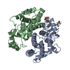
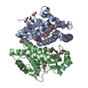
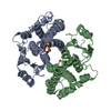
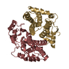
- Assembly
| Deposited unit | 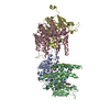
| ||||||||||||||||||||
|---|---|---|---|---|---|---|---|---|---|---|---|---|---|---|---|---|---|---|---|---|---|
| 1 | 
| ||||||||||||||||||||
| 2 | 
| ||||||||||||||||||||
| Unit cell |
| ||||||||||||||||||||
| Noncrystallographic symmetry (NCS) | NCS oper:
|
-Components
| #1: Protein | Mass: 26926.604 Da / Num. of mol.: 4 Source method: isolated from a genetically manipulated source Source: (gene. exp.) Sinorhizobium meliloti (bacteria) / Gene: gst2, SM2011_c00097 / Production host: #2: Chemical | ChemComp-GSH /   Mass: 307.323 Da / Num. of mol.: 4 / Source method: obtained synthetically / Formula: C10H17N3O6S Mass: 307.323 Da / Num. of mol.: 4 / Source method: obtained synthetically / Formula: C10H17N3O6S#3: Water | ChemComp-HOH / |  Mass: 18.015 Da / Num. of mol.: 229 / Source method: isolated from a natural source / Formula: H2O Mass: 18.015 Da / Num. of mol.: 229 / Source method: isolated from a natural source / Formula: H2O |
|---|
-Experimental details
-Experiment
| Experiment | Method: X-RAY DIFFRACTION / Number of used crystals: 1 |
|---|
- Sample preparation
| Crystal | Density Matthews: 2.32 Å3/Da / Density % sol: 46.98 % |
|---|---|
| Crystal grow | pH: 6.5 Details: Protein in 10 mM HEPES, pH 7.5, 150 mM sodium chloride, 5% glycerol, reservoir: 25% PEG3350, 0.1M bis-tris pH 6.5, 0.2M ammonium acetate, 5 mM GSH, cryoprotectant: none, vapor diffusion, ...Details: Protein in 10 mM HEPES, pH 7.5, 150 mM sodium chloride, 5% glycerol, reservoir: 25% PEG3350, 0.1M bis-tris pH 6.5, 0.2M ammonium acetate, 5 mM GSH, cryoprotectant: none, vapor diffusion, sitting drop, temperature 298K |
-Data collection
| Diffraction | Mean temperature: 100 K |
|---|---|
| Diffraction source | Source: SYNCHROTRON / Site: NSLS  / Beamline: X29A / Wavelength: 1.075 / Beamline: X29A / Wavelength: 1.075 |
| Detector | Type: ADSC QUANTUM 315 / Detector: CCD / Date: Oct 17, 2013 / Details: MIRRORS |
| Radiation | Monochromator: ROSENBAUM-ROCK DOUBLE CRYSTAL / Protocol: SINGLE WAVELENGTH / Monochromatic (M) / Laue (L): M / Scattering type: x-ray |
| Radiation wavelength | Wavelength: 1.075 Å / Relative weight: 1 |
| Reflection | Resolution: 2→50 Å / Num. obs: 62866 / % possible obs: 96.1 % / Observed criterion σ(I): -5 / Redundancy: 2 % / Rsym value: 0.047 / Net I/σ(I): 10.6 |
| Reflection shell | Resolution: 2→2.03 Å / Redundancy: 1.8 % / Rmerge(I) obs: 0.6 / Mean I/σ(I) obs: 1.1 / % possible all: 83.6 |
- Processing
| Software |
| ||||||||||||||||||||||||||||||||||||||||||||||||||||||||||||||||||||||||||||||||||||||||||||||||||||||||||||||||||||||||||||||||||||||||||||||||||||||||||||||||||||||||||||||||||||||
|---|---|---|---|---|---|---|---|---|---|---|---|---|---|---|---|---|---|---|---|---|---|---|---|---|---|---|---|---|---|---|---|---|---|---|---|---|---|---|---|---|---|---|---|---|---|---|---|---|---|---|---|---|---|---|---|---|---|---|---|---|---|---|---|---|---|---|---|---|---|---|---|---|---|---|---|---|---|---|---|---|---|---|---|---|---|---|---|---|---|---|---|---|---|---|---|---|---|---|---|---|---|---|---|---|---|---|---|---|---|---|---|---|---|---|---|---|---|---|---|---|---|---|---|---|---|---|---|---|---|---|---|---|---|---|---|---|---|---|---|---|---|---|---|---|---|---|---|---|---|---|---|---|---|---|---|---|---|---|---|---|---|---|---|---|---|---|---|---|---|---|---|---|---|---|---|---|---|---|---|---|---|---|---|
| Refinement | Method to determine structure: MOLECULAR REPLACEMENT Starting model: PDB ENTRY 3LSZ Resolution: 2→50 Å / Cor.coef. Fo:Fc: 0.974 / Cor.coef. Fo:Fc free: 0.95 / SU B: 13.136 / SU ML: 0.162 / Cross valid method: THROUGHOUT / ESU R: 0.165 / ESU R Free: 0.157 / Stereochemistry target values: MAXIMUM LIKELIHOOD / Details: HYDROGENS HAVE BEEN ADDED IN THE RIDING POSITIONS
| ||||||||||||||||||||||||||||||||||||||||||||||||||||||||||||||||||||||||||||||||||||||||||||||||||||||||||||||||||||||||||||||||||||||||||||||||||||||||||||||||||||||||||||||||||||||
| Solvent computation | Ion probe radii: 0.8 Å / Shrinkage radii: 0.8 Å / VDW probe radii: 1.2 Å / Solvent model: MASK | ||||||||||||||||||||||||||||||||||||||||||||||||||||||||||||||||||||||||||||||||||||||||||||||||||||||||||||||||||||||||||||||||||||||||||||||||||||||||||||||||||||||||||||||||||||||
| Displacement parameters | Biso mean: 62.183 Å2
| ||||||||||||||||||||||||||||||||||||||||||||||||||||||||||||||||||||||||||||||||||||||||||||||||||||||||||||||||||||||||||||||||||||||||||||||||||||||||||||||||||||||||||||||||||||||
| Refinement step | Cycle: LAST / Resolution: 2→50 Å
| ||||||||||||||||||||||||||||||||||||||||||||||||||||||||||||||||||||||||||||||||||||||||||||||||||||||||||||||||||||||||||||||||||||||||||||||||||||||||||||||||||||||||||||||||||||||
| Refine LS restraints |
|