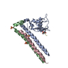
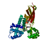
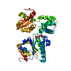
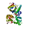
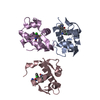
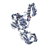
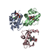
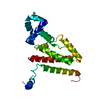
[English] 日本語
Yorodumi
- PDB-4nbx: Crystal Structure of Clostridium difficile Toxin A fragment TcdA-... -

+
Open data


ID or keywords:

Loading...

-
Basic information

Entry
Database: PDB / ID: 4nbx
TitleCrystal Structure of Clostridium difficile Toxin A fragment TcdA-A1 Bound to A20.1 VHH
Components
  • A20.1 VHH
  • TcdA
KeywordsIMMUNE SYSTEM / Antibody-antigen complex
Function / homology
Function and homology information


host cell cytosol / Transferases; Glycosyltransferases; Hexosyltransferases / glycosyltransferase activity / cysteine-type peptidase activity / host cell endosome membrane / toxin activity / Hydrolases; Acting on peptide bonds (peptidases); Cysteine endopeptidases / lipid binding / host cell plasma membrane / proteolysis ...host cell cytosol / Transferases; Glycosyltransferases; Hexosyltransferases / glycosyltransferase activity / cysteine-type peptidase activity / host cell endosome membrane / toxin activity / Hydrolases; Acting on peptide bonds (peptidases); Cysteine endopeptidases / lipid binding / host cell plasma membrane / proteolysis / extracellular region / membrane / metal ion binding / plasma membrane
Similarity search - Function
Cholin Binding / left handed beta-beta-3-solenoid / TcdA/TcdB toxin, N-terminal helical domain / TcdB toxin N-terminal helical domain / TcdA/TcdB toxin, catalytic glycosyltransferase domain / TcdA/TcdB catalytic glycosyltransferase domain / TcdA/TcdB toxin, pore forming domain / TcdA/TcdB pore forming domain / CGT/MARTX, cysteine protease (CPD) domain / CGT/MARTX, cysteine protease (CPD) domain superfamily ...Cholin Binding / left handed beta-beta-3-solenoid / TcdA/TcdB toxin, N-terminal helical domain / TcdB toxin N-terminal helical domain / TcdA/TcdB toxin, catalytic glycosyltransferase domain / TcdA/TcdB catalytic glycosyltransferase domain / TcdA/TcdB toxin, pore forming domain / TcdA/TcdB pore forming domain / CGT/MARTX, cysteine protease (CPD) domain / CGT/MARTX, cysteine protease (CPD) domain superfamily / Peptidase C80 family / CGT/MARTX cysteine protease (CPD) domain profile. / Choline-binding repeat / Putative cell wall binding repeat / Cell wall/choline-binding repeat / Cell wall-binding repeat profile. / Nucleotide-diphospho-sugar transferases / Ribbon / Immunoglobulins / Immunoglobulin-like / Sandwich / Mainly Beta
Similarity search - Domain/homology
Biological speciesClostridium difficile (bacteria)
Lama glama (llama)
MethodX-RAY DIFFRACTION / SYNCHROTRON / MOLECULAR REPLACEMENT / Resolution: 1.75 Å
AuthorsMurase, T. / Eugenio, L. / Schorr, M. / Hussack, G. / Tanha, J. / Kitova, E.N. / Klassen, J.S. / Ng, K.K.S.
CitationJournal: J.Biol.Chem. / Year: 2014
Title: Structural Basis for Antibody Recognition in the Receptor-binding Domains of Toxins A and B from Clostridium difficile.
Authors: Murase, T. / Eugenio, L. / Schorr, M. / Hussack, G. / Tanha, J. / Kitova, E.N. / Klassen, J.S. / Ng, K.K.
History
DepositionOct 23, 2013Deposition site: RCSB / Processing site: RCSB
Revision 1.0Dec 11, 2013Provider: repository / Type: Initial release
Revision 1.1Jan 1, 2014Group: Database references
Revision 1.2Feb 12, 2014Group: Database references

-
Structure visualization

Structure viewerMolecule:
MolmilJmol/JSmol

Downloads & links

-
Assembly

Deposited unit
A: TcdA
B: A20.1 VHH


Theoretical massNumber of molelcules
Total (without water)32,9852
Polymers32,9852
Non-polymers00
Water4,450247
1


  • Idetical with deposited unit
  • defined by author
TypeNameSymmetry operationNumber
identity operation1_555x,y,z1
Unit cell
Length a, b, c (Å)96.970, 96.970, 61.120
Angle α, β, γ (deg.)90.00, 90.00, 120.00
Int Tables number170
Space group name H-MP65

-
Components

#1: Protein TcdA


Mass: 16156.988 Da / Num. of mol.: 1 / Fragment: UNP residues 2573-2709
Source method: isolated from a genetically manipulated source
Source: (gene. exp.) Clostridium difficile (bacteria) / Strain: 48489 / Gene: tcdA / Production host: Escherichia coli (E. coli) / References: UniProt: I6YE93, UniProt: P16154*PLUS
#2: Antibody A20.1 VHH


Mass: 16828.449 Da / Num. of mol.: 1
Source method: isolated from a genetically manipulated source
Source: (gene. exp.) Lama glama (llama) / Production host: Escherichia coli (E. coli)
#3: Water ChemComp-HOH / water


Mass: 18.015 Da / Num. of mol.: 247 / Source method: isolated from a natural source / Formula: H2O

-
Experimental details

-
Experiment

ExperimentMethod: X-RAY DIFFRACTION / Number of used crystals: 1

-
Sample preparation

CrystalDensity Matthews: 2.51 Å3/Da / Density % sol: 51.09 %
Crystal growTemperature: 295 K / Method: vapor diffusion, hanging drop / pH: 7.5
Details: 0.6 M K/Na Tartrate, 0.1 M Tris-Cl, pH 7.5, 0.4 M TMAO, VAPOR DIFFUSION, HANGING DROP, temperature 295K

-
Data collection

DiffractionMean temperature: 100 K
Diffraction sourceSource: SYNCHROTRON / Site: CLSI / Beamline: 08B1-1 / Wavelength: 0.98 Å
DetectorType: MARMOSAIC 300 mm CCD / Detector: CCD / Date: Mar 9, 2010 / Details: mirrors
RadiationMonochromator: Si(111), double crystal / Protocol: SINGLE WAVELENGTH / Monochromatic (M) / Laue (L): M / Scattering type: x-ray
Radiation wavelengthWavelength: 0.98 Å / Relative weight: 1
ReflectionResolution: 1.75→40 Å / Num. all: 30256 / Num. obs: 30256 / % possible obs: 96.1 % / Observed criterion σ(F): -3 / Observed criterion σ(I): -3 / Redundancy: 4.3 % / Biso Wilson estimate: 27.2 Å2 / Rmerge(I) obs: 0.029 / Rsym value: 0.029 / Net I/σ(I): 30.9
Reflection shellResolution: 1.75→1.81 Å / Redundancy: 4.2 % / Rmerge(I) obs: 0.154 / Mean I/σ(I) obs: 8.9 / Num. unique all: 2068 / Rsym value: 0.154 / % possible all: 87.7

-
Processing

Software
NameVersionClassification
MxDCdata collection
PHASERphasing
REFMAC5.7.0029refinement
XDSdata reduction
XSCALEdata scaling
RefinementMethod to determine structure: MOLECULAR REPLACEMENT / Resolution: 1.75→31.74 Å / Cor.coef. Fo:Fc: 0.95 / Cor.coef. Fo:Fc free: 0.93 / SU B: 2.13 / SU ML: 0.071 / Cross valid method: THROUGHOUT / σ(F): 0 / σ(I): 0 / ESU R: 0.118 / ESU R Free: 0.115 / Stereochemistry target values: Engh & Huber
RfactorNum. reflection% reflectionSelection details
Rfree0.2287 1593 5 %RANDOM
Rwork0.19542 ---
obs0.19708 30256 96.1 %-
all-30256 --
Solvent computationIon probe radii: 0.8 Å / Shrinkage radii: 0.8 Å / VDW probe radii: 1.4 Å / Solvent model: BABINET MODEL WITH MASK
Displacement parametersBiso mean: 26.706 Å2
Baniso -1Baniso -2Baniso -3
1-0.28 Å20.28 Å20 Å2
2--0.28 Å20 Å2
3----0.91 Å2
Refinement stepCycle: LAST / Resolution: 1.75→31.74 Å
ProteinNucleic acidLigandSolventTotal
Num. atoms2091 0 0 247 2338
Refine LS restraints
Refine-IDTypeDev idealDev ideal targetNumber
X-RAY DIFFRACTIONr_bond_refined_d0.0060.0192155
X-RAY DIFFRACTIONr_bond_other_d
X-RAY DIFFRACTIONr_angle_refined_deg0.9691.912925
X-RAY DIFFRACTIONr_angle_other_deg
X-RAY DIFFRACTIONr_dihedral_angle_1_deg5.7935264
X-RAY DIFFRACTIONr_dihedral_angle_2_deg33.06323.304112
X-RAY DIFFRACTIONr_dihedral_angle_3_deg11.3415304
X-RAY DIFFRACTIONr_dihedral_angle_4_deg16.5681513
X-RAY DIFFRACTIONr_chiral_restr0.0690.2288
X-RAY DIFFRACTIONr_gen_planes_refined0.0040.0211737
X-RAY DIFFRACTIONr_gen_planes_other
X-RAY DIFFRACTIONr_nbd_refined
X-RAY DIFFRACTIONr_nbd_other
X-RAY DIFFRACTIONr_nbtor_refined
X-RAY DIFFRACTIONr_nbtor_other
X-RAY DIFFRACTIONr_xyhbond_nbd_refined
X-RAY DIFFRACTIONr_xyhbond_nbd_other
X-RAY DIFFRACTIONr_metal_ion_refined
X-RAY DIFFRACTIONr_metal_ion_other
X-RAY DIFFRACTIONr_symmetry_vdw_refined
X-RAY DIFFRACTIONr_symmetry_vdw_other
X-RAY DIFFRACTIONr_symmetry_hbond_refined
X-RAY DIFFRACTIONr_symmetry_hbond_other
X-RAY DIFFRACTIONr_symmetry_metal_ion_refined
X-RAY DIFFRACTIONr_symmetry_metal_ion_other
X-RAY DIFFRACTIONr_mcbond_it
X-RAY DIFFRACTIONr_mcbond_other
X-RAY DIFFRACTIONr_mcangle_it
X-RAY DIFFRACTIONr_mcangle_other
X-RAY DIFFRACTIONr_scbond_it
X-RAY DIFFRACTIONr_scbond_other
X-RAY DIFFRACTIONr_scangle_it
X-RAY DIFFRACTIONr_scangle_other
X-RAY DIFFRACTIONr_long_range_B_refined
X-RAY DIFFRACTIONr_long_range_B_other
X-RAY DIFFRACTIONr_rigid_bond_restr
X-RAY DIFFRACTIONr_sphericity_free
X-RAY DIFFRACTIONr_sphericity_bonded
LS refinement shellResolution: 1.75→1.795 Å / Total num. of bins used: 20
RfactorNum. reflection% reflection
Rfree0.293 97 -
Rwork0.224 1844 -
obs-1941 79.52 %

+
About Yorodumi

-
News

-
Feb 9, 2022. New format data for meta-information of EMDB entries

New format data for meta-information of EMDB entries

  • Version 3 of the EMDB header file is now the official format.
  • The previous official version 1.9 will be removed from the archive.

Related info.:EMDB header

External links:wwPDB to switch to version 3 of the EMDB data model

-
Aug 12, 2020. Covid-19 info

Covid-19 info

URL: https://pdbjlvh1.pdbj.org/emnavi/covid19.php

New page: Covid-19 featured information page in EM Navigator.

Related info.:Covid-19 info / Mar 5, 2020. Novel coronavirus structure data

+
Mar 5, 2020. Novel coronavirus structure data

Novel coronavirus structure data

Related info.:Yorodumi Speices / Aug 12, 2020. Covid-19 info

External links:COVID-19 featured content - PDBj / Molecule of the Month (242):Coronavirus Proteases

+
Jan 31, 2019. EMDB accession codes are about to change! (news from PDBe EMDB page)

EMDB accession codes are about to change! (news from PDBe EMDB page)

  • The allocation of 4 digits for EMDB accession codes will soon come to an end. Whilst these codes will remain in use, new EMDB accession codes will include an additional digit and will expand incrementally as the available range of codes is exhausted. The current 4-digit format prefixed with “EMD-” (i.e. EMD-XXXX) will advance to a 5-digit format (i.e. EMD-XXXXX), and so on. It is currently estimated that the 4-digit codes will be depleted around Spring 2019, at which point the 5-digit format will come into force.
  • The EM Navigator/Yorodumi systems omit the EMD- prefix.

Related info.:Q: What is EMD? / ID/Accession-code notation in Yorodumi/EM Navigator

External links:EMDB Accession Codes are Changing Soon! / Contact to PDBj

+
Jul 12, 2017. Major update of PDB

Major update of PDB

  • wwPDB released updated PDB data conforming to the new PDBx/mmCIF dictionary.
  • This is a major update changing the version number from 4 to 5, and with Remediation, in which all the entries are updated.
  • In this update, many items about electron microscopy experimental information are reorganized (e.g. em_software).
  • Now, EM Navigator and Yorodumi are based on the updated data.

External links:wwPDB Remediation / Enriched Model Files Conforming to OneDep Data Standards Now Available in the PDB FTP Archive

-
Yorodumi

Thousand views of thousand structures

  • Yorodumi is a browser for structure data from EMDB, PDB, SASBDB, etc.
  • This page is also the successor to EM Navigator detail page, and also detail information page/front-end page for Omokage search.
  • The word "yorodu" (or yorozu) is an old Japanese word meaning "ten thousand". "mi" (miru) is to see.

Related info.:EMDB / PDB / SASBDB / Comparison of 3 databanks / Yorodumi Search / Aug 31, 2016. New EM Navigator & Yorodumi / Yorodumi Papers / Jmol/JSmol / Function and homology information / Changes in new EM Navigator and Yorodumi

Read more