 Movie
Movie Controller

[English] 日本語
 Yorodumi
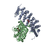
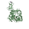
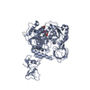
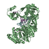
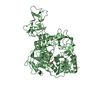
- PDB-4n0t: Core structure of the U6 small nuclear ribonucleoprotein at 1.7 A... -
+ Open data

- Basic information
| Entry | Database: PDB / ID: 4n0t | ||||||
|---|---|---|---|---|---|---|---|
| Title | Core structure of the U6 small nuclear ribonucleoprotein at 1.7 Angstrom resolution | ||||||
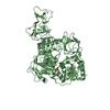
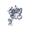
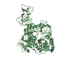
 Components Components |
| ||||||
 Keywords Keywords | RNA BINDING PROTEIN/RNA / spliceosomal ribonucleoprotein complex / annealing U6 and U4 snRNA / nucleus / RNA BINDING PROTEIN-RNA complex | ||||||
| Function / homology |  Function and homology information Function and homology informationU6 snRNP / snRNA binding / spliceosomal complex assembly / spliceosomal tri-snRNP complex assembly / U6 snRNA binding / spliceosomal complex / mRNA splicing, via spliceosome / ribonucleoprotein complex / RNA binding / nucleus Similarity search - Function | ||||||
| Biological species |  | ||||||
| Method |  X-RAY DIFFRACTION / SYNCHROTRON / MOLECULAR REPLACEMENT / Resolution: 1.7 Å X-RAY DIFFRACTION / SYNCHROTRON / MOLECULAR REPLACEMENT / Resolution: 1.7 Å | ||||||
 Authors Authors | Montemayor, E.J. / Curran, E.C. / Liao, H. / Andrews, K.L. / Treba, C.N. / Butcher, S.E. / Brow, D.A. | ||||||
 Citation Citation | Journal: Nat.Struct.Mol.Biol. / Year: 2014 Title: Core structure of the U6 small nuclear ribonucleoprotein at 1.7- angstrom resolution. Authors: Montemayor, E.J. / Curran, E.C. / Liao, H.H. / Andrews, K.L. / Treba, C.N. / Butcher, S.E. / Brow, D.A. | ||||||
| History |
|
- Structure visualization
| Structure viewer | Molecule: MolmilJmol/JSmol |
|---|
- Downloads & links
-Download
| PDBx/mmCIF format | 4n0t.cif.gz | 161.3 KB | Display | PDBx/mmCIF format |
|---|---|---|---|---|
| PDB format | pdb4n0t.ent.gz | 121.8 KB | Display | PDB format |
| PDBx/mmJSON format | 4n0t.json.gz | Tree view | PDBx/mmJSON format | |
| Others |  Other downloads Other downloads |
-Validation report
| Arichive directory | https://data.pdbj.org/pub/pdb/validation_reports/n0/4n0tftp://data.pdbj.org/pub/pdb/validation_reports/n0/4n0t | HTTPS FTP |
|---|
-Related structure data
| Related structure data | 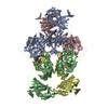 2ghpS S: Starting model for refinement |
|---|---|
| Similar structure data |


-Links
 PDBj
PDBj


- Assembly
| Deposited unit | 
| ||||||||
|---|---|---|---|---|---|---|---|---|---|
| 1 |
| ||||||||
| Unit cell |
|
-Components
| #1: Protein | Mass: 43246.758 Da / Num. of mol.: 1 Source method: isolated from a genetically manipulated source Source: (gene. exp.) Strain: S288c / Gene: PRP24, YM8156.10C, YMR268C / Plasmid: pET3a / Production host:  |
|---|---|
| #2: RNA chain | Mass: 23184.857 Da / Num. of mol.: 1 / Mutation: A62G, U100C, U101C / Source method: obtained synthetically Details: In vitro transcription using a cloned plasmid template References: GenBank: BK006945.2 |
| #3: Chemical | ChemComp-SO4 /   Mass: 96.063 Da / Num. of mol.: 1 / Source method: obtained synthetically / Formula: SO4 Mass: 96.063 Da / Num. of mol.: 1 / Source method: obtained synthetically / Formula: SO4 |
| #4: Water | ChemComp-HOH /  Mass: 18.015 Da / Num. of mol.: 696 / Source method: isolated from a natural source / Formula: H2O Mass: 18.015 Da / Num. of mol.: 696 / Source method: isolated from a natural source / Formula: H2O |
-Experimental details
-Experiment
| Experiment | Method: X-RAY DIFFRACTION / Number of used crystals: 1 |
|---|
- Sample preparation
| Crystal | Density Matthews: 2.57 Å3/Da / Density % sol: 52.06 % |
|---|---|
| Crystal grow | Temperature: 274 K / Method: vapor diffusion, sitting drop / pH: 5.5 Details: 100 mM lithium sulfate, 100 mM sodium citrate, pH 5.5, 20 % PEG 1,000, VAPOR DIFFUSION, SITTING DROP, temperature 274K |
-Data collection
| Diffraction | Mean temperature: 100 K |
|---|---|
| Diffraction source | Source: SYNCHROTRON / Site: APS  / Beamline: 21-ID-F / Wavelength: 0.97872 Å / Beamline: 21-ID-F / Wavelength: 0.97872 Å |
| Detector | Type: MARMOSAIC 225 mm CCD / Detector: CCD / Date: Jun 15, 2013 |
| Radiation | Monochromator: diamond laue monochromator / Protocol: SINGLE WAVELENGTH / Monochromatic (M) / Laue (L): M / Scattering type: x-ray |
| Radiation wavelength | Wavelength: 0.97872 Å / Relative weight: 1 |
| Reflection | Resolution: 1.7→50 Å / % possible obs: 99.6 % / Observed criterion σ(F): 0 / Observed criterion σ(I): 0 / Net I/σ(I): 15.3 |
| Reflection shell | Resolution: 1.7→1.73 Å / Redundancy: 7.5 % / Mean I/σ(I) obs: 2.5 / Rsym value: 0.9 / % possible all: 97 |
- Processing
| Software |
| |||||||||||||||||||||||||||||||||||||||||||||||||||||||||||||||||||||||||||||||||||||||||||||||||||||||||||||||||||||||||||||||||||||||||||||||||||||||||||||||||||||||||||||||||||||||||||||||||||||||||||||||||||||||||
|---|---|---|---|---|---|---|---|---|---|---|---|---|---|---|---|---|---|---|---|---|---|---|---|---|---|---|---|---|---|---|---|---|---|---|---|---|---|---|---|---|---|---|---|---|---|---|---|---|---|---|---|---|---|---|---|---|---|---|---|---|---|---|---|---|---|---|---|---|---|---|---|---|---|---|---|---|---|---|---|---|---|---|---|---|---|---|---|---|---|---|---|---|---|---|---|---|---|---|---|---|---|---|---|---|---|---|---|---|---|---|---|---|---|---|---|---|---|---|---|---|---|---|---|---|---|---|---|---|---|---|---|---|---|---|---|---|---|---|---|---|---|---|---|---|---|---|---|---|---|---|---|---|---|---|---|---|---|---|---|---|---|---|---|---|---|---|---|---|---|---|---|---|---|---|---|---|---|---|---|---|---|---|---|---|---|---|---|---|---|---|---|---|---|---|---|---|---|---|---|---|---|---|---|---|---|---|---|---|---|---|---|---|---|---|---|---|---|---|
| Refinement | Method to determine structure: MOLECULAR REPLACEMENT Starting model: PDB ENTRY 2GHP Resolution: 1.7→26.25 Å / SU ML: 0.18 / Cross valid method: THROUGHOUT / σ(F): 1.34 / Phase error: 23.21 / Stereochemistry target values: ML
| |||||||||||||||||||||||||||||||||||||||||||||||||||||||||||||||||||||||||||||||||||||||||||||||||||||||||||||||||||||||||||||||||||||||||||||||||||||||||||||||||||||||||||||||||||||||||||||||||||||||||||||||||||||||||
| Solvent computation | Shrinkage radii: 0.9 Å / VDW probe radii: 1.11 Å / Solvent model: FLAT BULK SOLVENT MODEL | |||||||||||||||||||||||||||||||||||||||||||||||||||||||||||||||||||||||||||||||||||||||||||||||||||||||||||||||||||||||||||||||||||||||||||||||||||||||||||||||||||||||||||||||||||||||||||||||||||||||||||||||||||||||||
| Refinement step | Cycle: LAST / Resolution: 1.7→26.25 Å
| |||||||||||||||||||||||||||||||||||||||||||||||||||||||||||||||||||||||||||||||||||||||||||||||||||||||||||||||||||||||||||||||||||||||||||||||||||||||||||||||||||||||||||||||||||||||||||||||||||||||||||||||||||||||||
| Refine LS restraints |
| |||||||||||||||||||||||||||||||||||||||||||||||||||||||||||||||||||||||||||||||||||||||||||||||||||||||||||||||||||||||||||||||||||||||||||||||||||||||||||||||||||||||||||||||||||||||||||||||||||||||||||||||||||||||||
| LS refinement shell |
| |||||||||||||||||||||||||||||||||||||||||||||||||||||||||||||||||||||||||||||||||||||||||||||||||||||||||||||||||||||||||||||||||||||||||||||||||||||||||||||||||||||||||||||||||||||||||||||||||||||||||||||||||||||||||
| Refinement TLS params. | Method: refined / Refine-ID: X-RAY DIFFRACTION
| |||||||||||||||||||||||||||||||||||||||||||||||||||||||||||||||||||||||||||||||||||||||||||||||||||||||||||||||||||||||||||||||||||||||||||||||||||||||||||||||||||||||||||||||||||||||||||||||||||||||||||||||||||||||||
| Refinement TLS group |
|
