 Movie
Movie Controller

[English] 日本語
 Yorodumi
- PDB-4n0o: Complex structure of Arterivirus nonstructural protein 10 (helica... -
+ Open data

- Basic information
| Entry | Database: PDB / ID: 4n0o | ||||||
|---|---|---|---|---|---|---|---|
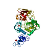
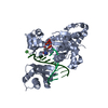
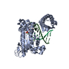
| Title | Complex structure of Arterivirus nonstructural protein 10 (helicase) with DNA | ||||||
 Components Components |
| ||||||
 Keywords Keywords | HYDROLASE/DNA / arterivirus / helicase / zbd / nsp10 / complex / HYDROLASE-DNA complex | ||||||
| Function / homology |  Function and homology information Function and homology informationserine-type exopeptidase activity / Hydrolases; Acting on peptide bonds (peptidases); Serine endopeptidases / host cell membrane / RNA nuclease activity / DNA helicase activity / Lyases; Phosphorus-oxygen lyases / endonuclease activity / symbiont-mediated suppression of host ISG15-protein conjugation / DNA helicase / symbiont-mediated perturbation of host ubiquitin-like protein modification ...serine-type exopeptidase activity / Hydrolases; Acting on peptide bonds (peptidases); Serine endopeptidases / host cell membrane / RNA nuclease activity / DNA helicase activity / Lyases; Phosphorus-oxygen lyases / endonuclease activity / symbiont-mediated suppression of host ISG15-protein conjugation / DNA helicase / symbiont-mediated perturbation of host ubiquitin-like protein modification / ubiquitinyl hydrolase 1 / cysteine-type deubiquitinase activity / Hydrolases; Acting on peptide bonds (peptidases); Cysteine endopeptidases / RNA helicase activity / lyase activity / viral protein processing / host cell perinuclear region of cytoplasm / RNA helicase / symbiont-mediated suppression of host type I interferon-mediated signaling pathway / serine-type endopeptidase activity / viral translational frameshifting / RNA-directed RNA polymerase / cysteine-type endopeptidase activity / viral RNA genome replication / RNA-directed RNA polymerase activity / DNA-templated transcription / host cell nucleus / ATP hydrolysis activity / proteolysis / RNA binding / zinc ion binding / ATP binding Similarity search - Function | ||||||
| Biological species |  Equine arteritis virus Equine arteritis virus | ||||||
| Method |  X-RAY DIFFRACTION / SYNCHROTRON / MOLECULAR REPLACEMENT / Resolution: 2.65 Å X-RAY DIFFRACTION / SYNCHROTRON / MOLECULAR REPLACEMENT / Resolution: 2.65 Å | ||||||
 Authors Authors | Deng, Z. / Chen, Z. | ||||||
 Citation Citation | Journal: Nucleic Acids Res. / Year: 2014 Title: Structural basis for the regulatory function of a complex zinc-binding domain in a replicative arterivirus helicase resembling a nonsense-mediated mRNA decay helicase. Authors: Deng, Z. / Lehmann, K.C. / Li, X. / Feng, C. / Wang, G. / Zhang, Q. / Qi, X. / Yu, L. / Zhang, X. / Feng, W. / Wu, W. / Gong, P. / Tao, Y. / Posthuma, C.C. / Snijder, E.J. / Gorbalenya, A.E. / Chen, Z. | ||||||
| History |
|
- Structure visualization
| Structure viewer | Molecule: MolmilJmol/JSmol |
|---|
- Downloads & links
-Download
| PDBx/mmCIF format | 4n0o.cif.gz | 334.9 KB | Display | PDBx/mmCIF format |
|---|---|---|---|---|
| PDB format | pdb4n0o.ent.gz | 266.3 KB | Display | PDB format |
| PDBx/mmJSON format | 4n0o.json.gz | Tree view | PDBx/mmJSON format | |
| Others |  Other downloads Other downloads |
-Validation report
| Arichive directory | https://data.pdbj.org/pub/pdb/validation_reports/n0/4n0oftp://data.pdbj.org/pub/pdb/validation_reports/n0/4n0o | HTTPS FTP |
|---|
-Related structure data
| Related structure data |  4n0nSC S: Starting model for refinement C: citing same article ( |
|---|---|
| Similar structure data |


-Links
 PDBj
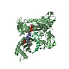
PDBj- Assembly
| Deposited unit | 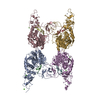
| ||||||||
|---|---|---|---|---|---|---|---|---|---|
| 1 | 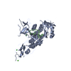
| ||||||||
| 2 | 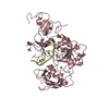
| ||||||||
| 3 | 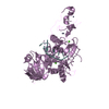
| ||||||||
| 4 | 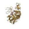
| ||||||||
| Unit cell |
|
-Components
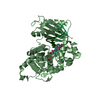
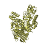
| #1: Protein | Mass: 45655.383 Da / Num. of mol.: 4 Source method: isolated from a genetically manipulated source Source: (gene. exp.) Equine arteritis virus / Strain: equine arteritis virus / Gene: 1a-1b, nsp10, rep / Plasmid: pet-28a / Production host:  References: UniProt: P19811, Hydrolases; Acting on peptide bonds (peptidases); Cysteine endopeptidases, ubiquitinyl hydrolase 1, Hydrolases; Acting on peptide bonds (peptidases); Serine ...References: UniProt: P19811, Hydrolases; Acting on peptide bonds (peptidases); Cysteine endopeptidases, ubiquitinyl hydrolase 1, Hydrolases; Acting on peptide bonds (peptidases); Serine endopeptidases, RNA-directed RNA polymerase, DNA helicase, RNA helicase #2: DNA chain | Mass: 2084.392 Da / Num. of mol.: 4 / Source method: obtained synthetically #3: Chemical | ChemComp-ZN /   Mass: 65.409 Da / Num. of mol.: 12 / Source method: obtained synthetically / Formula: Zn Mass: 65.409 Da / Num. of mol.: 12 / Source method: obtained synthetically / Formula: Zn#4: Chemical | ChemComp-CA /   Mass: 40.078 Da / Num. of mol.: 7 / Source method: obtained synthetically / Formula: Ca Mass: 40.078 Da / Num. of mol.: 7 / Source method: obtained synthetically / Formula: Ca#5: Water | ChemComp-HOH / |  Mass: 18.015 Da / Num. of mol.: 833 / Source method: isolated from a natural source / Formula: H2O Mass: 18.015 Da / Num. of mol.: 833 / Source method: isolated from a natural source / Formula: H2O |
|---|
-Experimental details
-Experiment
| Experiment | Method: X-RAY DIFFRACTION / Number of used crystals: 3 |
|---|
- Sample preparation
| Crystal | Density Matthews: 3.18 Å3/Da / Density % sol: 61.3 % |
|---|---|
| Crystal grow | Temperature: 277 K / Method: vapor diffusion, hanging drop / pH: 7 Details: 1.2M NH4SO4, pH 7, VAPOR DIFFUSION, HANGING DROP, temperature 277K |
-Data collection
| Diffraction | Mean temperature: 130 K | |||||||||||||||
|---|---|---|---|---|---|---|---|---|---|---|---|---|---|---|---|---|
| Diffraction source | Source: SYNCHROTRON / Site: SSRF  / Beamline: BL17U / Wavelength: 1 Å / Beamline: BL17U / Wavelength: 1 Å | |||||||||||||||
| Detector | Type: ADSC QUANTUM 270 / Detector: CCD / Date: Mar 4, 2012 | |||||||||||||||
| Radiation | Monochromator: unknown / Protocol: SINGLE WAVELENGTH / Monochromatic (M) / Laue (L): M / Scattering type: x-ray | |||||||||||||||
| Radiation wavelength | Wavelength: 1 Å / Relative weight: 1 | |||||||||||||||
| Reflection twin |
| |||||||||||||||
| Reflection | Resolution: 2.65→50 Å / Num. all: 69690 / Num. obs: 66954 / % possible obs: 96.1 % / Observed criterion σ(F): 3 / Observed criterion σ(I): 3 | |||||||||||||||
| Reflection shell | Resolution: 2.65→2.7 Å / % possible all: 95.3 |
- Processing
| Software |
| ||||||||||||||||||||||||||||||||||||||||||||||||||||||||||||||||||||||||||||||||||||||||||||||||||||||||||||||||||||||||||||||||||||||||||||||||||||||||||||||||||||||||||||||||||||||
|---|---|---|---|---|---|---|---|---|---|---|---|---|---|---|---|---|---|---|---|---|---|---|---|---|---|---|---|---|---|---|---|---|---|---|---|---|---|---|---|---|---|---|---|---|---|---|---|---|---|---|---|---|---|---|---|---|---|---|---|---|---|---|---|---|---|---|---|---|---|---|---|---|---|---|---|---|---|---|---|---|---|---|---|---|---|---|---|---|---|---|---|---|---|---|---|---|---|---|---|---|---|---|---|---|---|---|---|---|---|---|---|---|---|---|---|---|---|---|---|---|---|---|---|---|---|---|---|---|---|---|---|---|---|---|---|---|---|---|---|---|---|---|---|---|---|---|---|---|---|---|---|---|---|---|---|---|---|---|---|---|---|---|---|---|---|---|---|---|---|---|---|---|---|---|---|---|---|---|---|---|---|---|---|
| Refinement | Method to determine structure: MOLECULAR REPLACEMENT Starting model: PDB ENTRY 4N0N Resolution: 2.65→50 Å / Cor.coef. Fo:Fc: 0.896 / Cor.coef. Fo:Fc free: 0.89 / SU B: 7.358 / SU ML: 0.167 / Cross valid method: THROUGHOUT / σ(F): 3 / ESU R: 0.16 / ESU R Free: 0.07 / Stereochemistry target values: MAXIMUM LIKELIHOOD / Details: HYDROGENS HAVE BEEN USED IF PRESENT IN THE INPUT
| ||||||||||||||||||||||||||||||||||||||||||||||||||||||||||||||||||||||||||||||||||||||||||||||||||||||||||||||||||||||||||||||||||||||||||||||||||||||||||||||||||||||||||||||||||||||
| Solvent computation | Ion probe radii: 0.8 Å / Shrinkage radii: 0.8 Å / VDW probe radii: 1.2 Å / Solvent model: MASK | ||||||||||||||||||||||||||||||||||||||||||||||||||||||||||||||||||||||||||||||||||||||||||||||||||||||||||||||||||||||||||||||||||||||||||||||||||||||||||||||||||||||||||||||||||||||
| Displacement parameters | Biso mean: 48.364 Å2
| ||||||||||||||||||||||||||||||||||||||||||||||||||||||||||||||||||||||||||||||||||||||||||||||||||||||||||||||||||||||||||||||||||||||||||||||||||||||||||||||||||||||||||||||||||||||
| Refinement step | Cycle: LAST / Resolution: 2.65→50 Å
| ||||||||||||||||||||||||||||||||||||||||||||||||||||||||||||||||||||||||||||||||||||||||||||||||||||||||||||||||||||||||||||||||||||||||||||||||||||||||||||||||||||||||||||||||||||||
| Refine LS restraints |
|