 Movie
Movie Controller

[English] 日本語
 Yorodumi
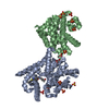
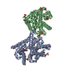
- PDB-4muw: Crystal Structure of PDE10A with Novel Keto-Benzimidazole Inhibitor -
+ Open data

- Basic information
| Entry | Database: PDB / ID: 4muw | ||||||
|---|---|---|---|---|---|---|---|
| Title | Crystal Structure of PDE10A with Novel Keto-Benzimidazole Inhibitor | ||||||
 Components Components | cAMP and cAMP-inhibited cGMP 3',5'-cyclic phosphodiesterase 10A | ||||||
 Keywords Keywords | HYDROLASE/HYDROLASE INHIBITOR / PDE10A / phosphodiesterase 10A / inhibitors / Keto-Benzimidazoles / HYDROLASE-HYDROLASE INHIBITOR complex | ||||||
| Function / homology |  Function and homology information Function and homology information3',5'-cGMP-stimulated cyclic-nucleotide phosphodiesterase activity / 3',5'-cyclic-nucleotide phosphodiesterase / negative regulation of receptor guanylyl cyclase signaling pathway / cGMP catabolic process / cAMP catabolic process / cGMP effects / 3',5'-cyclic-nucleotide phosphodiesterase activity / cGMP binding / 3',5'-cyclic-GMP phosphodiesterase activity / 3',5'-cyclic-AMP phosphodiesterase activity ...3',5'-cGMP-stimulated cyclic-nucleotide phosphodiesterase activity / 3',5'-cyclic-nucleotide phosphodiesterase / negative regulation of receptor guanylyl cyclase signaling pathway / cGMP catabolic process / cAMP catabolic process / cGMP effects / 3',5'-cyclic-nucleotide phosphodiesterase activity / cGMP binding / 3',5'-cyclic-GMP phosphodiesterase activity / 3',5'-cyclic-AMP phosphodiesterase activity / regulation of adenylate cyclase-activating G protein-coupled receptor signaling pathway / cAMP binding / negative regulation of cAMP/PKA signal transduction / G alpha (s) signalling events / glutamatergic synapse / signal transduction / metal ion binding / cytosol Similarity search - Function | ||||||
| Biological species |  Homo sapiens (human) Homo sapiens (human) | ||||||
| Method |  X-RAY DIFFRACTION / MOLECULAR REPLACEMENT / Resolution: 2.639 Å X-RAY DIFFRACTION / MOLECULAR REPLACEMENT / Resolution: 2.639 Å | ||||||
 Authors Authors | Chmait, S. / Jordan, S. | ||||||
 Citation Citation | Journal: J.Med.Chem. / Year: 2013 Title: Design, Optimization, and Biological Evaluation of Novel Keto-Benzimidazoles as Potent and Selective Inhibitors of Phosphodiesterase 10A (PDE10A). Authors: Hu, E. / Kunz, R.K. / Chen, N. / Rumfelt, S. / Siegmund, A. / Andrews, K. / Chmait, S. / Zhao, S. / Davis, C. / Chen, H. / Lester-Zeiner, D. / Ma, J. / Biorn, C. / Shi, J. / Porter, A. / ...Authors: Hu, E. / Kunz, R.K. / Chen, N. / Rumfelt, S. / Siegmund, A. / Andrews, K. / Chmait, S. / Zhao, S. / Davis, C. / Chen, H. / Lester-Zeiner, D. / Ma, J. / Biorn, C. / Shi, J. / Porter, A. / Treanor, J. / Allen, J.R. | ||||||
| History |
|
- Structure visualization
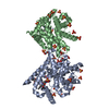
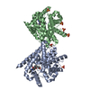
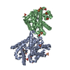
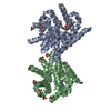
| Structure viewer | Molecule: MolmilJmol/JSmol |
|---|
- Downloads & links
-Download
| PDBx/mmCIF format | 4muw.cif.gz | 270.8 KB | Display | PDBx/mmCIF format |
|---|---|---|---|---|
| PDB format | pdb4muw.ent.gz | 220.3 KB | Display | PDB format |
| PDBx/mmJSON format | 4muw.json.gz | Tree view | PDBx/mmJSON format | |
| Others |  Other downloads Other downloads |
-Validation report
| Arichive directory | https://data.pdbj.org/pub/pdb/validation_reports/mu/4muwftp://data.pdbj.org/pub/pdb/validation_reports/mu/4muw | HTTPS FTP |
|---|
-Related structure data
| Related structure data |  4mvhC  4ddlS S: Starting model for refinement C: citing same article ( |
|---|---|
| Similar structure data |
-Links
 PDBj
PDBj

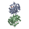
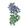
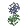
- Assembly
| Deposited unit | 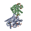
| ||||||||
|---|---|---|---|---|---|---|---|---|---|
| 1 |
| ||||||||
| 2 | 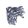
| ||||||||
| 3 | 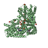
| ||||||||
| Unit cell |
|
-Components
-Protein , 1 types, 2 molecules AB
| #1: Protein | Mass: 40575.398 Da / Num. of mol.: 2 / Fragment: human PDE10a, residues 442-779 Source method: isolated from a genetically manipulated source Source: (gene. exp.) Homo sapiens (human) / Gene: PDE10A / Production host:  References: UniProt: Q9Y233, 3',5'-cyclic-nucleotide phosphodiesterase, 3',5'-cyclic-GMP phosphodiesterase |
|---|
-Non-polymers , 5 types, 189 molecules 


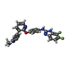

| #2: Chemical | ChemComp-ZN /  Mass: 65.409 Da / Num. of mol.: 4 / Source method: obtained synthetically / Formula: Zn Mass: 65.409 Da / Num. of mol.: 4 / Source method: obtained synthetically / Formula: Zn#3: Chemical | ChemComp-SO4 /  Mass: 96.063 Da / Num. of mol.: 15 / Source method: obtained synthetically / Formula: SO4 Mass: 96.063 Da / Num. of mol.: 15 / Source method: obtained synthetically / Formula: SO4#4: Chemical | ChemComp-GOL / |  Mass: 92.094 Da / Num. of mol.: 1 / Source method: obtained synthetically / Formula: C3H8O3 Mass: 92.094 Da / Num. of mol.: 1 / Source method: obtained synthetically / Formula: C3H8O3#5: Chemical |  Mass: 444.436 Da / Num. of mol.: 2 / Source method: obtained synthetically / Formula: C24H18F2N6O Mass: 444.436 Da / Num. of mol.: 2 / Source method: obtained synthetically / Formula: C24H18F2N6O#6: Water | ChemComp-HOH / | Mass: 18.015 Da / Num. of mol.: 167 / Source method: isolated from a natural source / Formula: H2O |
|---|
-Details
| Has protein modification | Y |
|---|
-Experimental details
-Experiment
| Experiment | Method: X-RAY DIFFRACTION / Number of used crystals: 1 |
|---|
- Sample preparation
| Crystal | Density Matthews: 4.14 Å3/Da / Density % sol: 70.29 % |
|---|---|
| Crystal grow | Temperature: 277 K / Method: vapor diffusion, hanging drop / pH: 6.5 Details: 1.6M Ammonium Sulfate, 0.1M MES monohydrate, 10% v/v 1,4-Dioxane, pH 6.5, VAPOR DIFFUSION, HANGING DROP, temperature 277K |
-Data collection
| Diffraction | Mean temperature: 100 K |
|---|---|
| Diffraction source | Source: ROTATING ANODE / Type: RIGAKU FR-E SUPERBRIGHT / Wavelength: 1.5418 Å |
| Detector | Type: RIGAKU SATURN 92 / Detector: CCD / Date: Jan 7, 2013 / Details: Osmic Varimax HF |
| Radiation | Protocol: SINGLE WAVELENGTH / Monochromatic (M) / Laue (L): M / Scattering type: x-ray |
| Radiation wavelength | Wavelength: 1.5418 Å / Relative weight: 1 |
| Reflection | Resolution: 2.639→29.19 Å / Num. all: 39231 / Num. obs: 39179 / % possible obs: 100 % / Observed criterion σ(F): 0 / Observed criterion σ(I): 0 |
| Reflection shell | Resolution: 2.639→2.69 Å / % possible all: 99.8 |
- Processing
| Software |
| |||||||||||||||||||||||||||||||||||||||||||||||||||||||||||||||||||||||||||||||||||||||||||||||||||||||||
|---|---|---|---|---|---|---|---|---|---|---|---|---|---|---|---|---|---|---|---|---|---|---|---|---|---|---|---|---|---|---|---|---|---|---|---|---|---|---|---|---|---|---|---|---|---|---|---|---|---|---|---|---|---|---|---|---|---|---|---|---|---|---|---|---|---|---|---|---|---|---|---|---|---|---|---|---|---|---|---|---|---|---|---|---|---|---|---|---|---|---|---|---|---|---|---|---|---|---|---|---|---|---|---|---|---|---|
| Refinement | Method to determine structure: MOLECULAR REPLACEMENT Starting model: PDB ENTRY 4DDL Resolution: 2.639→29.19 Å / Cor.coef. Fo:Fc: 0.965 / Cor.coef. Fo:Fc free: 0.956 / SU B: 13.627 / SU ML: 0.142 / Cross valid method: THROUGHOUT / ESU R: 0.271 / ESU R Free: 0.201 Stereochemistry target values: MAXIMUM LIKELIHOOD WITH PHASES Details: HYDROGENS HAVE BEEN ADDED IN THE RIDING POSITIONS
| |||||||||||||||||||||||||||||||||||||||||||||||||||||||||||||||||||||||||||||||||||||||||||||||||||||||||
| Solvent computation | Ion probe radii: 0.8 Å / Shrinkage radii: 0.8 Å / VDW probe radii: 1.2 Å / Solvent model: MASK | |||||||||||||||||||||||||||||||||||||||||||||||||||||||||||||||||||||||||||||||||||||||||||||||||||||||||
| Displacement parameters | Biso mean: 50.92 Å2
| |||||||||||||||||||||||||||||||||||||||||||||||||||||||||||||||||||||||||||||||||||||||||||||||||||||||||
| Refinement step | Cycle: LAST / Resolution: 2.639→29.19 Å
| |||||||||||||||||||||||||||||||||||||||||||||||||||||||||||||||||||||||||||||||||||||||||||||||||||||||||
| Refine LS restraints |
| |||||||||||||||||||||||||||||||||||||||||||||||||||||||||||||||||||||||||||||||||||||||||||||||||||||||||
| LS refinement shell | Resolution: 2.639→2.707 Å / Total num. of bins used: 20
| |||||||||||||||||||||||||||||||||||||||||||||||||||||||||||||||||||||||||||||||||||||||||||||||||||||||||
| Refinement TLS params. | Method: refined / Refine-ID: X-RAY DIFFRACTION
| |||||||||||||||||||||||||||||||||||||||||||||||||||||||||||||||||||||||||||||||||||||||||||||||||||||||||
| Refinement TLS group |
|
