 Movie
Movie Controller

[English] 日本語
 Yorodumi
- PDB-4mrg: Crystal structure of the murine cd44 hyaluronan binding domain co... -
+ Open data

- Basic information
| Entry | Database: PDB / ID: 4mrg | ||||||
|---|---|---|---|---|---|---|---|
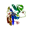
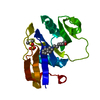
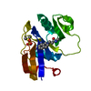
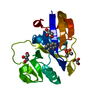
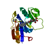
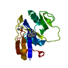
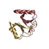
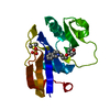
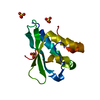
| Title | Crystal structure of the murine cd44 hyaluronan binding domain complex with a small molecule | ||||||
 Components Components | CD44 antigen | ||||||
 Keywords Keywords | Cell adhesion/inhibitor / Link module / Cell receptor / Hyaluronan binding / Cell surface / Cell adhesion-inhibitor complex | ||||||
| Function / homology |  Function and homology information Function and homology informationHyaluronan metabolism / Hyaluronan degradation / positive regulation of monocyte aggregation / macrophage fusion / hyaluronic acid binding / macrophage migration inhibitory factor receptor complex / Degradation of the extracellular matrix / Integrin cell surface interactions / regulation of lamellipodium morphogenesis / postsynapse organization ...Hyaluronan metabolism / Hyaluronan degradation / positive regulation of monocyte aggregation / macrophage fusion / hyaluronic acid binding / macrophage migration inhibitory factor receptor complex / Degradation of the extracellular matrix / Integrin cell surface interactions / regulation of lamellipodium morphogenesis / postsynapse organization / Cell surface interactions at the vascular wall / monocyte aggregation / wound healing involved in inflammatory response / hyaluronan catabolic process / branching involved in prostate gland morphogenesis / positive regulation of adaptive immune response / type II transforming growth factor beta receptor binding / negative regulation of CD4-positive, alpha-beta T cell proliferation / negative regulation of mature B cell apoptotic process / positive regulation of neutrophil apoptotic process / regulation of modification of postsynaptic structure / channel regulator activity / positive regulation of heterotypic cell-cell adhesion / wound healing, spreading of cells / epidermal growth factor receptor binding / cargo receptor activity / branching involved in ureteric bud morphogenesis / negative regulation of regulatory T cell differentiation / negative regulation of intrinsic apoptotic signaling pathway in response to DNA damage by p53 class mediator / negative regulation of DNA damage response, signal transduction by p53 class mediator / microvillus / lamellipodium membrane / cell projection / Neutrophil degranulation / cellular response to fibroblast growth factor stimulus / receptor-mediated endocytosis / T cell activation / phosphoprotein binding / regulation of cell growth / negative regulation of inflammatory response / Wnt signaling pathway / neuron projection development / cytokine-mediated signaling pathway / transmembrane signaling receptor activity / cell migration / presynapse / basolateral plasma membrane / positive regulation of ERK1 and ERK2 cascade / cell adhesion / postsynapse / apical plasma membrane / membrane raft / inflammatory response / external side of plasma membrane / positive regulation of gene expression / protein kinase binding / negative regulation of apoptotic process / glutamatergic synapse / Golgi apparatus / cell surface / protein-containing complex / extracellular region / plasma membrane / cytosol Similarity search - Function | ||||||
| Biological species |  | ||||||
| Method |  X-RAY DIFFRACTION / SYNCHROTRON / MOLECULAR REPLACEMENT / molecular replacement / Resolution: 1.69 Å X-RAY DIFFRACTION / SYNCHROTRON / MOLECULAR REPLACEMENT / molecular replacement / Resolution: 1.69 Å | ||||||
 Authors Authors | Liu, L.K. / Finzel, B. | ||||||
 Citation Citation | Journal: J.Med.Chem. / Year: 2014 Title: Fragment-Based Identification of an Inducible Binding Site on Cell Surface Receptor CD44 for the Design of Protein-Carbohydrate Interaction Inhibitors. Authors: Liu, L.K. / Finzel, B.C. #1: Journal: Nat.Struct.Mol.Biol. / Year: 2007Title: Structures of the Cd44-hyaluronan complex provide insight into a fundamental carbohydrate-protein interaction. Authors: Banerji, S. / Wright, A.J. / Noble, M. / Mahoney, D.J. / Campbell, I.D. / Day, A.J. / Jackson, D.G. | ||||||
| History |
|
- Structure visualization
| Structure viewer | Molecule: MolmilJmol/JSmol |
|---|
- Downloads & links
-Download
| PDBx/mmCIF format | 4mrg.cif.gz | 47.6 KB | Display | PDBx/mmCIF format |
|---|---|---|---|---|
| PDB format | pdb4mrg.ent.gz | 32.6 KB | Display | PDB format |
| PDBx/mmJSON format | 4mrg.json.gz | Tree view | PDBx/mmJSON format | |
| Others |  Other downloads Other downloads |
-Validation report
| Arichive directory | https://data.pdbj.org/pub/pdb/validation_reports/mr/4mrgftp://data.pdbj.org/pub/pdb/validation_reports/mr/4mrg | HTTPS FTP |
|---|
-Related structure data
| Related structure data |  4mrdC  4mreC 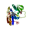 4mrfC 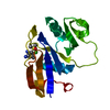 4mrhC  4np2C  4np3C  2jcpS C: citing same article ( S: Starting model for refinement |
|---|---|
| Similar structure data |

-Links
 PDBj
PDBj


- Assembly
| Deposited unit | 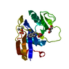
| ||||||||
|---|---|---|---|---|---|---|---|---|---|
| 1 |
| ||||||||
| Unit cell |
|
-Components
-Protein , 1 types, 1 molecules A
| #1: Protein | Mass: 16724.604 Da / Num. of mol.: 1 / Fragment: HYALURONAN BINDING DOMAIN, RESIDUES 23-174 / Mutation: H23M, Q24N Source method: isolated from a genetically manipulated source Source: (gene. exp.)  |
|---|
-Non-polymers , 5 types, 90 molecules 


| #2: Chemical | ChemComp-24W /  Mass: 148.205 Da / Num. of mol.: 1 / Source method: obtained synthetically / Formula: C9H12N2 Mass: 148.205 Da / Num. of mol.: 1 / Source method: obtained synthetically / Formula: C9H12N2 | ||||
|---|---|---|---|---|---|
| #3: Chemical | ChemComp-DMS /  Mass: 78.133 Da / Num. of mol.: 1 / Source method: obtained synthetically / Formula: C2H6OS / Comment: DMSO, precipitant*YM Mass: 78.133 Da / Num. of mol.: 1 / Source method: obtained synthetically / Formula: C2H6OS / Comment: DMSO, precipitant*YM | ||||
| #4: Chemical |  Mass: 92.094 Da / Num. of mol.: 2 / Source method: obtained synthetically / Formula: C3H8O3 Mass: 92.094 Da / Num. of mol.: 2 / Source method: obtained synthetically / Formula: C3H8O3#5: Chemical | ChemComp-SO4 / |  Mass: 96.063 Da / Num. of mol.: 1 / Source method: obtained synthetically / Formula: SO4 Mass: 96.063 Da / Num. of mol.: 1 / Source method: obtained synthetically / Formula: SO4#6: Water | ChemComp-HOH / | Mass: 18.015 Da / Num. of mol.: 85 / Source method: isolated from a natural source / Formula: H2O |
-Details
| Has protein modification | Y |
|---|
-Experimental details
-Experiment
| Experiment | Method: X-RAY DIFFRACTION / Number of used crystals: 1 |
|---|
- Sample preparation
| Crystal | Density Matthews: 2.14 Å3/Da / Density % sol: 42.41 % |
|---|---|
| Crystal grow | Temperature: 298 K / Method: vapor diffusion, hanging drop / pH: 6.5 Details: 30% PEG MME 5000, 100 mM MES, 200 mM (NH4)2SO4, pH 6.5, VAPOR DIFFUSION, HANGING DROP, temperature 298K |
-Data collection
| Diffraction | Mean temperature: 100 K |
|---|---|
| Diffraction source | Source: SYNCHROTRON / Site: ALS  / Beamline: 4.2.2 / Wavelength: 1 Å / Beamline: 4.2.2 / Wavelength: 1 Å |
| Detector | Type: NOIR-1 / Detector: CCD / Date: Dec 16, 2012 |
| Radiation | Monochromator: Rosenbaum-Rock monochromator Si 111 / Protocol: SINGLE WAVELENGTH / Monochromatic (M) / Laue (L): M / Scattering type: x-ray |
| Radiation wavelength | Wavelength: 1 Å / Relative weight: 1 |
| Reflection | Resolution: 1.69→40.8 Å / Num. all: 56490 / Num. obs: 56490 / % possible obs: 95.7 % / Redundancy: 3.7 % / Biso Wilson estimate: 18.4 Å2 / Rmerge(I) obs: 0.039 / Rsym value: 0.046 / Net I/σ(I): 27.2 |
| Reflection shell | Resolution: 1.69→1.73 Å / Redundancy: 2.7 % / Rmerge(I) obs: 0.197 / Mean I/σ(I) obs: 5.3 / Num. unique all: 871 / Rsym value: 0.243 / % possible all: 74.8 |
-Phasing
| Phasing | Method: molecular replacement | |||||||||
|---|---|---|---|---|---|---|---|---|---|---|
| Phasing MR | Rfactor: 32.85 / Model details: Phaser MODE: MR_AUTO
|
- Processing
| Software |
| |||||||||||||||||||||||||||||||||||||||||||||||||||||||||||||||||
|---|---|---|---|---|---|---|---|---|---|---|---|---|---|---|---|---|---|---|---|---|---|---|---|---|---|---|---|---|---|---|---|---|---|---|---|---|---|---|---|---|---|---|---|---|---|---|---|---|---|---|---|---|---|---|---|---|---|---|---|---|---|---|---|---|---|---|
| Refinement | Method to determine structure: MOLECULAR REPLACEMENT Starting model: pdb entry 2JCP Resolution: 1.69→40.8 Å / Cor.coef. Fo:Fc: 0.955 / Cor.coef. Fo:Fc free: 0.919 / WRfactor Rfree: 0.1951 / WRfactor Rwork: 0.1536 / Occupancy max: 1 / Occupancy min: 0.5 / FOM work R set: 0.8898 / SU B: 1.811 / SU ML: 0.062 / SU R Cruickshank DPI: 0.1081 / SU Rfree: 0.1098 / Cross valid method: THROUGHOUT / ESU R: 0.108 / ESU R Free: 0.11 / Stereochemistry target values: MAXIMUM LIKELIHOOD Details: HYDROGENS HAVE BEEN ADDED IN THE RIDING POSITIONS U VALUES : REFINED INDIVIDUALLY
| |||||||||||||||||||||||||||||||||||||||||||||||||||||||||||||||||
| Solvent computation | Ion probe radii: 0.8 Å / Shrinkage radii: 0.8 Å / VDW probe radii: 1.4 Å / Solvent model: MASK | |||||||||||||||||||||||||||||||||||||||||||||||||||||||||||||||||
| Displacement parameters | Biso max: 43.54 Å2 / Biso mean: 10.718 Å2 / Biso min: 2.87 Å2
| |||||||||||||||||||||||||||||||||||||||||||||||||||||||||||||||||
| Refinement step | Cycle: LAST / Resolution: 1.69→40.8 Å
| |||||||||||||||||||||||||||||||||||||||||||||||||||||||||||||||||
| Refine LS restraints |
| |||||||||||||||||||||||||||||||||||||||||||||||||||||||||||||||||
| LS refinement shell | Resolution: 1.691→1.735 Å / Total num. of bins used: 20
|
