 Movie
Movie Controller

[English] 日本語
 Yorodumi
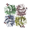
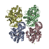
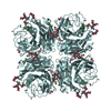
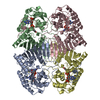
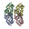
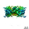
- PDB-4moz: Fructose-bisphosphate aldolase from Slackia heliotrinireducens DS... -
+ Open data

- Basic information
| Entry | Database: PDB / ID: 4moz | ||||||
|---|---|---|---|---|---|---|---|
| Title | Fructose-bisphosphate aldolase from Slackia heliotrinireducens DSM 20476 | ||||||
 Components Components | Fructose-bisphosphate aldolase | ||||||
 Keywords Keywords | LYASE / PSI-Biology / MCSG / Midwest Center for Structural Genomics / fructose-bisphosphate aldolase | ||||||
| Function / homology |  Function and homology information Function and homology informationfructose-bisphosphate aldolase / fructose-bisphosphate aldolase activity Similarity search - Function | ||||||
| Biological species |  Slackia heliotrinireducens (bacteria) Slackia heliotrinireducens (bacteria) | ||||||
| Method |  X-RAY DIFFRACTION / SYNCHROTRON / SAD / Resolution: 2.15 Å X-RAY DIFFRACTION / SYNCHROTRON / SAD / Resolution: 2.15 Å | ||||||
 Authors Authors | Chang, C. / Li, H. / Jedrzejczak, R. / Joachimiak, A. / Midwest Center for Structural Genomics (MCSG) | ||||||
 Citation Citation | Journal: To be Published Title: Fructose-bisphosphate aldolase from Slackia heliotrinireducens DSM 20476 Authors: Chang, C. / Li, H. / Jedrzejczak, R. / Joachimiak, A. | ||||||
| History |
|
- Structure visualization
| Structure viewer | Molecule: MolmilJmol/JSmol |
|---|
- Downloads & links
-Download
| PDBx/mmCIF format | 4moz.cif.gz | 630 KB | Display | PDBx/mmCIF format |
|---|---|---|---|---|
| PDB format | pdb4moz.ent.gz | 526.9 KB | Display | PDB format |
| PDBx/mmJSON format | 4moz.json.gz | Tree view | PDBx/mmJSON format | |
| Others |  Other downloads Other downloads |
-Validation report
| Arichive directory | https://data.pdbj.org/pub/pdb/validation_reports/mo/4mozftp://data.pdbj.org/pub/pdb/validation_reports/mo/4moz | HTTPS FTP |
|---|
-Related structure data
| Similar structure data | |
|---|---|
| Other databases |


-Links
 PDBj
PDBj
- Assembly
| Deposited unit | 
| ||||||||
|---|---|---|---|---|---|---|---|---|---|
| 1 |
| ||||||||
| 2 | 
| ||||||||
| Unit cell |
|
-Components
| #1: Protein | Mass: 34602.820 Da / Num. of mol.: 5 Source method: isolated from a genetically manipulated source Source: (gene. exp.) Slackia heliotrinireducens (bacteria) / Strain: DSM 20476 / Gene: Shel_19790 / Production host: #2: Water | ChemComp-HOH / |  Mass: 18.015 Da / Num. of mol.: 1114 / Source method: isolated from a natural source / Formula: H2O Mass: 18.015 Da / Num. of mol.: 1114 / Source method: isolated from a natural source / Formula: H2OHas protein modification | Y | |
|---|
-Experimental details
-Experiment
| Experiment | Method: X-RAY DIFFRACTION / Number of used crystals: 1 |
|---|
- Sample preparation
| Crystal | Density Matthews: 3.47 Å3/Da / Density % sol: 64.55 % |
|---|---|
| Crystal grow | Temperature: 289 K / Method: vapor diffusion, sitting drop / pH: 7.5 Details: 90 mM HEPES, 1.26M Sodium Citrate, 10% Glycerol, pH 7.5, VAPOR DIFFUSION, SITTING DROP, temperature 289K |
-Data collection
| Diffraction | Mean temperature: 100 K |
|---|---|
| Diffraction source | Source: SYNCHROTRON / Site: APS  / Beamline: 19-ID / Wavelength: 0.97923 Å / Beamline: 19-ID / Wavelength: 0.97923 Å |
| Detector | Type: ADSC QUANTUM 315r / Detector: CCD / Date: Oct 11, 2012 |
| Radiation | Monochromator: Si(111) double crystal / Protocol: SINGLE WAVELENGTH / Monochromatic (M) / Laue (L): M / Scattering type: x-ray |
| Radiation wavelength | Wavelength: 0.97923 Å / Relative weight: 1 |
| Reflection | Resolution: 2.15→50 Å / Num. all: 128812 / Num. obs: 128506 / % possible obs: 99.8 % / Observed criterion σ(I): -3 / Redundancy: 5.1 % / Rmerge(I) obs: 0.121 / Net I/σ(I): 18 |
| Reflection shell | Resolution: 2.15→2.17 Å / Redundancy: 4.8 % / Rmerge(I) obs: 0.765 / Mean I/σ(I) obs: 2.2 / Num. unique all: 3136 / % possible all: 99.5 |
- Processing
| Software |
| ||||||||||||||||||||||||||||||||||||||||||||||||||||||||||||
|---|---|---|---|---|---|---|---|---|---|---|---|---|---|---|---|---|---|---|---|---|---|---|---|---|---|---|---|---|---|---|---|---|---|---|---|---|---|---|---|---|---|---|---|---|---|---|---|---|---|---|---|---|---|---|---|---|---|---|---|---|---|
| Refinement | Method to determine structure: SAD / Resolution: 2.15→50 Å / Cor.coef. Fo:Fc: 0.961 / Cor.coef. Fo:Fc free: 0.936 / Occupancy max: 1 / Occupancy min: 0 / SU B: 10.352 / SU ML: 0.115 / Cross valid method: THROUGHOUT / σ(F): 0 / ESU R: 0.744 / ESU R Free: 0.162 Stereochemistry target values: MAXIMUM LIKELIHOOD WITH PHASES Details: HYDROGENS HAVE BEEN USED IF PRESENT IN THE INPUT U VALUES : REFINED INDIVIDUALLY
| ||||||||||||||||||||||||||||||||||||||||||||||||||||||||||||
| Solvent computation | Ion probe radii: 0.8 Å / Shrinkage radii: 0.8 Å / VDW probe radii: 1.2 Å / Solvent model: MASK | ||||||||||||||||||||||||||||||||||||||||||||||||||||||||||||
| Displacement parameters | Biso max: 111.63 Å2 / Biso mean: 33.0525 Å2 / Biso min: 15.05 Å2
| ||||||||||||||||||||||||||||||||||||||||||||||||||||||||||||
| Refinement step | Cycle: LAST / Resolution: 2.15→50 Å
| ||||||||||||||||||||||||||||||||||||||||||||||||||||||||||||
| Refine LS restraints |
| ||||||||||||||||||||||||||||||||||||||||||||||||||||||||||||
| LS refinement shell | Resolution: 2.154→2.21 Å / Total num. of bins used: 20
|
