 Movie
Movie Controller

[English] 日本語
 Yorodumi
- PDB-4mh6: 2.8 Angstrom Crystal Structure of Type III Secretion Protein YscO... -
+ Open data

- Basic information
| Entry | Database: PDB / ID: 4mh6 | ||||||
|---|---|---|---|---|---|---|---|
| Title | 2.8 Angstrom Crystal Structure of Type III Secretion Protein YscO from Vibrio parahaemolyticus | ||||||
 Components Components | Putative type III secretion protein YscO | ||||||
 Keywords Keywords | UNKNOWN FUNCTION / Structural Genomics / Center for Structural Genomics of Infectious Diseases / CSGID | ||||||
| Function / homology | Type III secretion sytem,YscO / Type III secretion protein YscO / Helix Hairpins - #1700 / : / Helix Hairpins / Orthogonal Bundle / Mainly Alpha / PHOSPHATE ION / Putative type III secretion protein YscO Function and homology information Function and homology information | ||||||
| Biological species |   Vibrio parahaemolyticus (bacteria) Vibrio parahaemolyticus (bacteria) | ||||||
| Method |  X-RAY DIFFRACTION / SYNCHROTRON / SAD / Resolution: 2.8 Å X-RAY DIFFRACTION / SYNCHROTRON / SAD / Resolution: 2.8 Å | ||||||
 Authors Authors | Minasov, G. / Wawrzak, Z. / Shuvalova, L. / Winsor, J. / Dubrovska, I. / Grimshaw, S. / Kwon, K. / Anderson, W.F. / Center for Structural Genomics of Infectious Diseases (CSGID) | ||||||
 Citation Citation | Journal: TO BE PUBLISHED Title: 2.8 Angstrom Crystal Structure of Type III Secretion Protein YscO from Vibrio parahaemolyticus. Authors: Minasov, G. / Wawrzak, Z. / Shuvalova, L. / Winsor, J. / Dubrovska, I. / Grimshaw, S. / Kwon, K. / Anderson, W.F. | ||||||
| History |
|
- Structure visualization
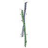
| Structure viewer | Molecule: MolmilJmol/JSmol |
|---|
- Downloads & links
-Download
| PDBx/mmCIF format | 4mh6.cif.gz | 85.5 KB | Display | PDBx/mmCIF format |
|---|---|---|---|---|
| PDB format | pdb4mh6.ent.gz | 66.3 KB | Display | PDB format |
| PDBx/mmJSON format | 4mh6.json.gz | Tree view | PDBx/mmJSON format | |
| Others |  Other downloads Other downloads |
-Validation report
| Arichive directory | https://data.pdbj.org/pub/pdb/validation_reports/mh/4mh6ftp://data.pdbj.org/pub/pdb/validation_reports/mh/4mh6 | HTTPS FTP |
|---|
-Related structure data
| Similar structure data | |
|---|---|
| Other databases |


-Links
 PDBj
PDBj- Assembly
| Deposited unit | 
| |||||||||
|---|---|---|---|---|---|---|---|---|---|---|
| 1 |
| |||||||||
| Unit cell |
| |||||||||
| Components on special symmetry positions |
|
-Components
| #1: Protein | Mass: 21322.688 Da / Num. of mol.: 1 Source method: isolated from a genetically manipulated source Source: (gene. exp.) Vibrio parahaemolyticus (bacteria) / Strain: RIMD 2210633 / Gene: VP1669, yscO / Plasmid: pMCSG7 / Production host:   Spodoptera frugiperda (fall armyworm) / Strain (production host): Rosetta gami 2 / References: UniProt: Q87P49 Spodoptera frugiperda (fall armyworm) / Strain (production host): Rosetta gami 2 / References: UniProt: Q87P49 | ||||
|---|---|---|---|---|---|
| #2: Chemical |   Mass: 94.971 Da / Num. of mol.: 2 / Source method: obtained synthetically / Formula: PO4 Mass: 94.971 Da / Num. of mol.: 2 / Source method: obtained synthetically / Formula: PO4#3: Water | ChemComp-HOH / |  Mass: 18.015 Da / Num. of mol.: 93 / Source method: isolated from a natural source / Formula: H2O Mass: 18.015 Da / Num. of mol.: 93 / Source method: isolated from a natural source / Formula: H2OHas protein modification | Y | |
-Experimental details
-Experiment
| Experiment | Method: X-RAY DIFFRACTION / Number of used crystals: 2 |
|---|
- Sample preparation
| Crystal | Density Matthews: 4.13 Å3/Da / Density % sol: 70.24 % |
|---|---|
| Crystal grow | Temperature: 295 K / Method: vapor diffusion, sitting drop / pH: 6.2 Details: Protein: 2.28 mG/mL, 0.5 M Sodium chloride, 0.01 M Tris-HCL buffer pH 8.3; Screen: JCSG+ (C9), 0.1M Sodium/Potasium phosphate pH 6.2, 25% (v/v) 1,2 Propanediol, 10% (v/v) Glycerol, VAPOR ...Details: Protein: 2.28 mG/mL, 0.5 M Sodium chloride, 0.01 M Tris-HCL buffer pH 8.3; Screen: JCSG+ (C9), 0.1M Sodium/Potasium phosphate pH 6.2, 25% (v/v) 1,2 Propanediol, 10% (v/v) Glycerol, VAPOR DIFFUSION, SITTING DROP, temperature 295K |
-Data collection
| Diffraction | Mean temperature: 100 K |
|---|---|
| Diffraction source | Source: SYNCHROTRON / Site: APS  / Beamline: 21-ID-F / Wavelength: 0.97872 Å / Beamline: 21-ID-F / Wavelength: 0.97872 Å |
| Detector | Type: MARMOSAIC 225 mm CCD / Detector: CCD / Date: Aug 20, 2013 / Details: Beryllium lenses |
| Radiation | Monochromator: Diamond / Protocol: SINGLE WAVELENGTH / Monochromatic (M) / Laue (L): M / Scattering type: x-ray |
| Radiation wavelength | Wavelength: 0.97872 Å / Relative weight: 1 |
| Reflection | Resolution: 2.8→30 Å / Num. all: 9084 / Num. obs: 9084 / % possible obs: 99.9 % / Observed criterion σ(I): -3 / Redundancy: 9.6 % / Biso Wilson estimate: 55.3 Å2 / Rmerge(I) obs: 0.139 / Net I/σ(I): 18.3 |
| Reflection shell | Resolution: 2.8→2.85 Å / Redundancy: 10 % / Rmerge(I) obs: 0.603 / Mean I/σ(I) obs: 4.2 / Num. unique all: 436 / % possible all: 100 |
- Processing
| Software |
| |||||||||||||||||||||||||||||||||||||||||||||||||||||||||||||||||||||||||||||||||||||||||||||||||||||||||||||||||||||||||||||||||||||||||||||||||||||||||||||||||||||||||||||||
|---|---|---|---|---|---|---|---|---|---|---|---|---|---|---|---|---|---|---|---|---|---|---|---|---|---|---|---|---|---|---|---|---|---|---|---|---|---|---|---|---|---|---|---|---|---|---|---|---|---|---|---|---|---|---|---|---|---|---|---|---|---|---|---|---|---|---|---|---|---|---|---|---|---|---|---|---|---|---|---|---|---|---|---|---|---|---|---|---|---|---|---|---|---|---|---|---|---|---|---|---|---|---|---|---|---|---|---|---|---|---|---|---|---|---|---|---|---|---|---|---|---|---|---|---|---|---|---|---|---|---|---|---|---|---|---|---|---|---|---|---|---|---|---|---|---|---|---|---|---|---|---|---|---|---|---|---|---|---|---|---|---|---|---|---|---|---|---|---|---|---|---|---|---|---|---|---|
| Refinement | Method to determine structure: SAD / Resolution: 2.8→29.24 Å / Cor.coef. Fo:Fc: 0.935 / Cor.coef. Fo:Fc free: 0.907 / SU B: 17.508 / SU ML: 0.187 Isotropic thermal model: Thermal Factors Individually Refined Cross valid method: THROUGHOUT / ESU R: 0.397 / ESU R Free: 0.286 / Stereochemistry target values: MAXIMUM LIKELIHOOD / Details: HYDROGENS HAVE BEEN ADDED IN THE RIDING POSITIONS
| |||||||||||||||||||||||||||||||||||||||||||||||||||||||||||||||||||||||||||||||||||||||||||||||||||||||||||||||||||||||||||||||||||||||||||||||||||||||||||||||||||||||||||||||
| Solvent computation | Ion probe radii: 0.8 Å / Shrinkage radii: 0.8 Å / VDW probe radii: 1.2 Å / Solvent model: MASK | |||||||||||||||||||||||||||||||||||||||||||||||||||||||||||||||||||||||||||||||||||||||||||||||||||||||||||||||||||||||||||||||||||||||||||||||||||||||||||||||||||||||||||||||
| Displacement parameters | Biso mean: 52.031 Å2
| |||||||||||||||||||||||||||||||||||||||||||||||||||||||||||||||||||||||||||||||||||||||||||||||||||||||||||||||||||||||||||||||||||||||||||||||||||||||||||||||||||||||||||||||
| Refinement step | Cycle: LAST / Resolution: 2.8→29.24 Å
| |||||||||||||||||||||||||||||||||||||||||||||||||||||||||||||||||||||||||||||||||||||||||||||||||||||||||||||||||||||||||||||||||||||||||||||||||||||||||||||||||||||||||||||||
| Refine LS restraints |
| |||||||||||||||||||||||||||||||||||||||||||||||||||||||||||||||||||||||||||||||||||||||||||||||||||||||||||||||||||||||||||||||||||||||||||||||||||||||||||||||||||||||||||||||
| LS refinement shell | Resolution: 2.8→2.872 Å / Total num. of bins used: 20
| |||||||||||||||||||||||||||||||||||||||||||||||||||||||||||||||||||||||||||||||||||||||||||||||||||||||||||||||||||||||||||||||||||||||||||||||||||||||||||||||||||||||||||||||
| Refinement TLS params. | Method: refined / Refine-ID: X-RAY DIFFRACTION
| |||||||||||||||||||||||||||||||||||||||||||||||||||||||||||||||||||||||||||||||||||||||||||||||||||||||||||||||||||||||||||||||||||||||||||||||||||||||||||||||||||||||||||||||
| Refinement TLS group |
|
