| Entry | Database: PDB / ID: 4mee
|
|---|
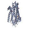
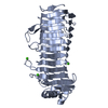
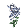
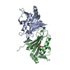
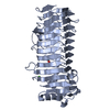
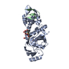
| Title | Crystal structure of the transport unit of the autotransporter AIDA-I from Escherichia coli |
|---|
 Components Components | Diffuse adherence adhesin |
|---|
 Keywords Keywords | PROTEIN BINDING / Beta barrel / outer membrane protein / Autotransporter |
|---|
| Function / homology |  Function and homology information Function and homology information
: / Adhesin of bacterial autotransporter system / Adhesin of bacterial autotransporter system, probable stalk / Autochaperone domain type 1 / Autochaperone Domain Type 1 / Autotransporter beta-domain / : / ESPR domain / Extended Signal Peptide of Type V secretion system / Autotransporter beta-domain ...: / Adhesin of bacterial autotransporter system / Adhesin of bacterial autotransporter system, probable stalk / Autochaperone domain type 1 / Autochaperone Domain Type 1 / Autotransporter beta-domain / : / ESPR domain / Extended Signal Peptide of Type V secretion system / Autotransporter beta-domain / Outer membrane autotransporter barrel / Autotransporter beta-domain / Autotransporter beta-domain profile. / Autotransporter beta-domain / Autotransporter beta-domain superfamily / Autotransporter, pectate lyase C-like domain superfamily / Pectin lyase fold/virulence factor / Lipocalin / Beta Barrel / Mainly BetaSimilarity search - Domain/homology |
|---|
| Biological species |   Escherichia coli (E. coli) Escherichia coli (E. coli) |
|---|
| Method |  X-RAY DIFFRACTION / SYNCHROTRON / MOLECULAR REPLACEMENT / Resolution: 3 Å X-RAY DIFFRACTION / SYNCHROTRON / MOLECULAR REPLACEMENT / Resolution: 3 Å |
|---|
 Authors Authors | Gawarzewski, I. / Tschapek, B. / Hoeppner, A. / Smits, S.H. / Jose, J. / Schmitt, L. |
|---|
 Citation Citation | Journal: J.Struct.Biol. / Year: 2014
Title: Crystal structure of the transport unit of the autotransporter adhesin involved in diffuse adherence from Escherichia coli.
Authors: Gawarzewski, I. / DiMaio, F. / Winterer, E. / Tschapek, B. / Smits, S.H. / Jose, J. / Schmitt, L. |
|---|
| History | | Deposition | Aug 26, 2013 | Deposition site: RCSB / Processing site: RCSB |
|---|
| Revision 1.0 | Jun 4, 2014 | Provider: repository / Type: Initial release |
|---|
| Revision 1.1 | Jul 23, 2014 | Group: Database references |
|---|
| Revision 1.2 | Feb 28, 2024 | Group: Data collection / Database references
Category: chem_comp_atom / chem_comp_bond ...chem_comp_atom / chem_comp_bond / database_2 / struct_ref_seq_dif
Item: _database_2.pdbx_DOI / _database_2.pdbx_database_accession / _struct_ref_seq_dif.details |
|---|
|
|---|
 Movie
Movie Controller

 Yorodumi
 Open data
 Basic information
 Structure visualization
 Downloads & links
 Other downloads
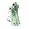


 PDBj
PDBj Assembly
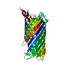
 Sample preparation
 / Beamline: ID23-2 / Wavelength: 0.8726 Å
 Processing