 Movie
Movie Controller


+ Open data

- Basic information
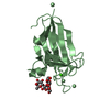
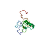
| Entry | Database: PDB / ID: 4m9v | |||||||||
|---|---|---|---|---|---|---|---|---|---|---|
| Title | Zfp57 mutant (E182Q) in complex with 5-carboxylcytosine DNA | |||||||||
 Components Components |
| |||||||||
 Keywords Keywords | TRANSCRIPTION/DNA / epigenetics / transcription factor / 5-carboxylcytosine / C2H2 Zinc finger / DNA binding / TRANSCRIPTION-DNA complex | |||||||||
| Function / homology |  Function and homology information Function and homology informationepigenetic programing of female pronucleus / autosome genomic imprinting / genomic imprinting / double-stranded methylated DNA binding / epigenetic programming in the zygotic pronuclei / negative regulation of gene expression via chromosomal CpG island methylation / heterochromatin / DNA-binding transcription repressor activity, RNA polymerase II-specific / RNA polymerase II cis-regulatory region sequence-specific DNA binding / chromatin binding ...epigenetic programing of female pronucleus / autosome genomic imprinting / genomic imprinting / double-stranded methylated DNA binding / epigenetic programming in the zygotic pronuclei / negative regulation of gene expression via chromosomal CpG island methylation / heterochromatin / DNA-binding transcription repressor activity, RNA polymerase II-specific / RNA polymerase II cis-regulatory region sequence-specific DNA binding / chromatin binding / negative regulation of transcription by RNA polymerase II / DNA-templated transcription / DNA binding / zinc ion binding / nucleus Similarity search - Function | |||||||||
| Biological species |  | |||||||||
| Method |  X-RAY DIFFRACTION / SYNCHROTRON / MOLECULAR REPLACEMENT / Resolution: 0.969 Å X-RAY DIFFRACTION / SYNCHROTRON / MOLECULAR REPLACEMENT / Resolution: 0.969 Å | |||||||||
 Authors Authors | Liu, Y. / Olanrewaju, Y.O. / Zhang, X. / Cheng, X. | |||||||||
 Citation Citation | Journal: Biochemistry / Year: 2013 Title: DNA recognition of 5-carboxylcytosine by a zfp57 mutant at an atomic resolution of 0.97 angstrom. Authors: Liu, Y. / Olanrewaju, Y.O. / Zhang, X. / Cheng, X. | |||||||||
| History |
|
- Structure visualization
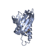
| Structure viewer | Molecule: MolmilJmol/JSmol |
|---|
- Downloads & links
-Download
| PDBx/mmCIF format | 4m9v.cif.gz | 178.1 KB | Display | PDBx/mmCIF format |
|---|---|---|---|---|
| PDB format | pdb4m9v.ent.gz | 139.8 KB | Display | PDB format |
| PDBx/mmJSON format | 4m9v.json.gz | Tree view | PDBx/mmJSON format | |
| Others |  Other downloads Other downloads |
-Validation report
| Arichive directory | https://data.pdbj.org/pub/pdb/validation_reports/m9/4m9vftp://data.pdbj.org/pub/pdb/validation_reports/m9/4m9v | HTTPS FTP |
|---|
-Related structure data
| Related structure data | 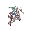 4gznS S: Starting model for refinement |
|---|---|
| Similar structure data |


-Links
 PDBj
PDBj


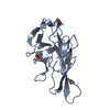
- Assembly
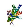
| Deposited unit | 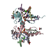
| ||||||||
|---|---|---|---|---|---|---|---|---|---|
| 1 | 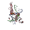
| ||||||||
| 2 | 
| ||||||||
| 3 |
| ||||||||
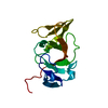
| Unit cell |
|
-Components
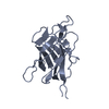
-DNA chain , 2 types, 4 molecules ADBE
| #1: DNA chain | Mass: 3363.224 Da / Num. of mol.: 2 / Source method: obtained synthetically / Details: DNA synthesis #2: DNA chain | Mass: 3402.221 Da / Num. of mol.: 2 / Source method: obtained synthetically / Details: DNA synthesis |
|---|
-Protein , 1 types, 2 molecules CF
| #3: Protein | Mass: 7376.424 Da / Num. of mol.: 2 / Mutation: E182Q Source method: isolated from a genetically manipulated source Source: (gene. exp.)  |
|---|
-Non-polymers , 5 types, 486 molecules 


| #4: Chemical | ChemComp-MPD / (  Mass: 118.174 Da / Num. of mol.: 4 / Source method: obtained synthetically / Formula: C6H14O2 / Comment: precipitant*YM Mass: 118.174 Da / Num. of mol.: 4 / Source method: obtained synthetically / Formula: C6H14O2 / Comment: precipitant*YM#5: Chemical | ChemComp-ACT /  Mass: 59.044 Da / Num. of mol.: 4 / Source method: obtained synthetically / Formula: C2H3O2 Mass: 59.044 Da / Num. of mol.: 4 / Source method: obtained synthetically / Formula: C2H3O2#6: Chemical |  Mass: 40.078 Da / Num. of mol.: 2 / Source method: obtained synthetically / Formula: Ca Mass: 40.078 Da / Num. of mol.: 2 / Source method: obtained synthetically / Formula: Ca#7: Chemical | ChemComp-ZN /  Mass: 65.409 Da / Num. of mol.: 4 / Source method: obtained synthetically / Formula: Zn Mass: 65.409 Da / Num. of mol.: 4 / Source method: obtained synthetically / Formula: Zn#8: Water | ChemComp-HOH / | Mass: 18.015 Da / Num. of mol.: 472 / Source method: isolated from a natural source / Formula: H2O |
|---|
-Experimental details
-Experiment
| Experiment | Method: X-RAY DIFFRACTION / Number of used crystals: 1 |
|---|
- Sample preparation
| Crystal | Density Matthews: 2.07 Å3/Da / Density % sol: 40.56 % |
|---|---|
| Crystal grow | Temperature: 289 K / Method: vapor diffusion, sitting drop / pH: 4.2 Details: 35% 2-methyl-2,4-pentanediol, 15% polyethylene glycol 8000, 100 mM CaCl2, and 100 mM acetate, pH 4.2, VAPOR DIFFUSION, SITTING DROP, temperature 289K |
-Data collection
| Diffraction | Mean temperature: 100 K |
|---|---|
| Diffraction source | Source: SYNCHROTRON / Site: APS  / Beamline: 22-ID / Wavelength: 0.8 Å / Beamline: 22-ID / Wavelength: 0.8 Å |
| Detector | Type: MARMOSAIC 300 mm CCD / Detector: CCD / Date: Dec 31, 2012 Details: Si 111. Rosenbaum-Rock double-crystal monochromator: liquid nitrogen cooled; sagitally focusing 2nd crystal, Rosenbaum-Rock vertical focusing mirror |
| Radiation | Monochromator: double crystal - liquid nitrogen cooled / Protocol: SINGLE WAVELENGTH / Monochromatic (M) / Laue (L): M / Scattering type: x-ray |
| Radiation wavelength | Wavelength: 0.8 Å / Relative weight: 1 |
| Reflection | Resolution: 0.969→33.5 Å / Num. obs: 129957 / % possible obs: 95.7 % / Observed criterion σ(I): -3 / Redundancy: 2.6 % / Biso Wilson estimate: 8.63 Å2 / Rmerge(I) obs: 0.07 / Net I/σ(I): 11.7 |
| Reflection shell | Resolution: 0.969→1 Å / Redundancy: 2.2 % / Rmerge(I) obs: 0.648 / Mean I/σ(I) obs: 1.4 / Num. unique all: 12599 / % possible all: 92.7 |
- Processing
| Software |
| |||||||||||||||||||||||||||||||||||||||||||||||||||||||||||||||||||||||||||||||||||||||||||||||||||||||||||||||||||||||||||||||||||||||||||||||||||||||||||||||||||||||||||||||||||||||||||||||||||||||||||||||||||||||||
|---|---|---|---|---|---|---|---|---|---|---|---|---|---|---|---|---|---|---|---|---|---|---|---|---|---|---|---|---|---|---|---|---|---|---|---|---|---|---|---|---|---|---|---|---|---|---|---|---|---|---|---|---|---|---|---|---|---|---|---|---|---|---|---|---|---|---|---|---|---|---|---|---|---|---|---|---|---|---|---|---|---|---|---|---|---|---|---|---|---|---|---|---|---|---|---|---|---|---|---|---|---|---|---|---|---|---|---|---|---|---|---|---|---|---|---|---|---|---|---|---|---|---|---|---|---|---|---|---|---|---|---|---|---|---|---|---|---|---|---|---|---|---|---|---|---|---|---|---|---|---|---|---|---|---|---|---|---|---|---|---|---|---|---|---|---|---|---|---|---|---|---|---|---|---|---|---|---|---|---|---|---|---|---|---|---|---|---|---|---|---|---|---|---|---|---|---|---|---|---|---|---|---|---|---|---|---|---|---|---|---|---|---|---|---|---|---|---|---|
| Refinement | Method to determine structure: MOLECULAR REPLACEMENT Starting model: PDB ENTRY 4GZN Resolution: 0.969→33.459 Å / SU ML: 0.08 / Cross valid method: THROUGHOUT / σ(F): 1.34 / Phase error: 14.11 / Stereochemistry target values: ML / Details: FLAT BULK SOLVENT MODEL
| |||||||||||||||||||||||||||||||||||||||||||||||||||||||||||||||||||||||||||||||||||||||||||||||||||||||||||||||||||||||||||||||||||||||||||||||||||||||||||||||||||||||||||||||||||||||||||||||||||||||||||||||||||||||||
| Solvent computation | Shrinkage radii: 0.9 Å / VDW probe radii: 1.11 Å / Solvent model: FLAT BULK SOLVENT MODEL | |||||||||||||||||||||||||||||||||||||||||||||||||||||||||||||||||||||||||||||||||||||||||||||||||||||||||||||||||||||||||||||||||||||||||||||||||||||||||||||||||||||||||||||||||||||||||||||||||||||||||||||||||||||||||
| Displacement parameters | Biso mean: 13.7 Å2 | |||||||||||||||||||||||||||||||||||||||||||||||||||||||||||||||||||||||||||||||||||||||||||||||||||||||||||||||||||||||||||||||||||||||||||||||||||||||||||||||||||||||||||||||||||||||||||||||||||||||||||||||||||||||||
| Refinement step | Cycle: LAST / Resolution: 0.969→33.459 Å
| |||||||||||||||||||||||||||||||||||||||||||||||||||||||||||||||||||||||||||||||||||||||||||||||||||||||||||||||||||||||||||||||||||||||||||||||||||||||||||||||||||||||||||||||||||||||||||||||||||||||||||||||||||||||||
| Refine LS restraints |
| |||||||||||||||||||||||||||||||||||||||||||||||||||||||||||||||||||||||||||||||||||||||||||||||||||||||||||||||||||||||||||||||||||||||||||||||||||||||||||||||||||||||||||||||||||||||||||||||||||||||||||||||||||||||||
| LS refinement shell | Refine-ID: X-RAY DIFFRACTION
|
