 Movie
Movie Controller

[English] 日本語
 Yorodumi
- PDB-4m6e: The high resolution structure of tyrocidine A reveals an amphipat... -
+ Open data

- Basic information
| Entry | Database: PDB / ID: 4m6e | ||||||
|---|---|---|---|---|---|---|---|
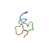
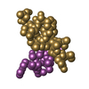
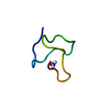
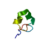
| Title | The high resolution structure of tyrocidine A reveals an amphipathic dimer | ||||||
 Components Components | tyrocidine A | ||||||
 Keywords Keywords | ANTIBIOTIC / cyclic peptide | ||||||
| Function / homology | tyrocidine A / METHANOL / :  Function and homology information Function and homology information | ||||||
| Biological species |  Bacillus brevis (bacteria) Bacillus brevis (bacteria) | ||||||
| Method |  X-RAY DIFFRACTION / SYNCHROTRON / AB INITIO PHASING / Resolution: 0.95 Å X-RAY DIFFRACTION / SYNCHROTRON / AB INITIO PHASING / Resolution: 0.95 Å | ||||||
 Authors Authors | Loll, P.J. / Economou, N.J. / Nahoum, V. | ||||||
 Citation Citation | Journal: Biochim.Biophys.Acta / Year: 2014 Title: The high resolution structure of tyrocidine A reveals an amphipathic dimer. Authors: Loll, P.J. / Upton, E.C. / Nahoum, V. / Economou, N.J. / Cocklin, S. | ||||||
| History |
|
- Structure visualization
| Structure viewer | Molecule: MolmilJmol/JSmol |
|---|
- Downloads & links
-Download
| PDBx/mmCIF format | 4m6e.cif.gz | 17.8 KB | Display | PDBx/mmCIF format |
|---|---|---|---|---|
| PDB format | pdb4m6e.ent.gz | 11.9 KB | Display | PDB format |
| PDBx/mmJSON format | 4m6e.json.gz | Tree view | PDBx/mmJSON format | |
| Others |  Other downloads Other downloads |
-Validation report
| Arichive directory | https://data.pdbj.org/pub/pdb/validation_reports/m6/4m6eftp://data.pdbj.org/pub/pdb/validation_reports/m6/4m6e | HTTPS FTP |
|---|
-Related structure data
| Similar structure data |
|---|


-Links
 PDBj
PDBj
- Assembly
| Deposited unit | 
| ||||||||||||
|---|---|---|---|---|---|---|---|---|---|---|---|---|---|
| 1 | 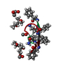
| ||||||||||||
| Unit cell |
| ||||||||||||
| Components on special symmetry positions |
|
-Components
| #1: Protein/peptide | 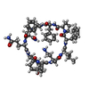  Type: Peptide-like / Class: Antibiotic / Mass: 1288.491 Da / Num. of mol.: 1 / Source method: isolated from a natural source / Source: (natural) Bacillus brevis (bacteria) / References: NOR: NOR00298, tyrocidine A Type: Peptide-like / Class: Antibiotic / Mass: 1288.491 Da / Num. of mol.: 1 / Source method: isolated from a natural source / Source: (natural) Bacillus brevis (bacteria) / References: NOR: NOR00298, tyrocidine A | ||||
|---|---|---|---|---|---|
| #2: Chemical | ChemComp-MPD / (  Mass: 118.174 Da / Num. of mol.: 1 / Source method: obtained synthetically / Formula: C6H14O2 / Comment: precipitant*YM Mass: 118.174 Da / Num. of mol.: 1 / Source method: obtained synthetically / Formula: C6H14O2 / Comment: precipitant*YM | ||||
| #3: Chemical |   Mass: 32.042 Da / Num. of mol.: 3 / Source method: obtained synthetically / Formula: CH4O Mass: 32.042 Da / Num. of mol.: 3 / Source method: obtained synthetically / Formula: CH4O#4: Water | ChemComp-HOH / |  Mass: 18.015 Da / Num. of mol.: 5 / Source method: isolated from a natural source / Formula: H2O Mass: 18.015 Da / Num. of mol.: 5 / Source method: isolated from a natural source / Formula: H2OHas protein modification | Y | |
-Experimental details
-Experiment
| Experiment | Method: X-RAY DIFFRACTION / Number of used crystals: 1 |
|---|
- Sample preparation
| Crystal | Density Matthews: 2.11 Å3/Da / Density % sol: 41.57 % |
|---|---|
| Crystal grow | Temperature: 291 K / Method: vapor diffusion, sitting drop Details: tyrocidine in methanol at 30 mg/mL, reservoir methanol:MPD (1:5 v/v), VAPOR DIFFUSION, SITTING DROP, temperature 291K |
-Data collection
| Diffraction | Mean temperature: 100 K |
|---|---|
| Diffraction source | Source: SYNCHROTRON / Site: NSLS  / Beamline: X6A / Wavelength: 0.8984 Å / Beamline: X6A / Wavelength: 0.8984 Å |
| Detector | Type: ADSC QUANTUM 270 / Detector: CCD / Date: Jan 1, 2008 |
| Radiation | Protocol: SINGLE WAVELENGTH / Monochromatic (M) / Laue (L): M / Scattering type: x-ray |
| Radiation wavelength | Wavelength: 0.8984 Å / Relative weight: 1 |
| Reflection | Resolution: 0.95→19 Å / Num. all: 6973 / Num. obs: 6973 / % possible obs: 98.8 % / Observed criterion σ(I): -3 / Redundancy: 12.2 % / Biso Wilson estimate: 9.6 Å2 / Rmerge(I) obs: 0.04 / Net I/σ(I): 48 |
- Processing
| Software |
| ||||||||||||||||||||||||||||||||||||||||||
|---|---|---|---|---|---|---|---|---|---|---|---|---|---|---|---|---|---|---|---|---|---|---|---|---|---|---|---|---|---|---|---|---|---|---|---|---|---|---|---|---|---|---|---|
| Refinement | Method to determine structure: AB INITIO PHASING / Resolution: 0.95→19 Å / SU ML: 0.05 / σ(F): 0 / Phase error: 16.57 / Stereochemistry target values: ML
| ||||||||||||||||||||||||||||||||||||||||||
| Solvent computation | Shrinkage radii: 0.9 Å / VDW probe radii: 1.11 Å / Solvent model: FLAT BULK SOLVENT MODEL | ||||||||||||||||||||||||||||||||||||||||||
| Refinement step | Cycle: LAST / Resolution: 0.95→19 Å
| ||||||||||||||||||||||||||||||||||||||||||
| Refine LS restraints |
| ||||||||||||||||||||||||||||||||||||||||||
| LS refinement shell |
|
