Resolution: 1.3→1.32 Å / Redundancy: 8.1 % / Rmerge(I) obs: 0.779 / Mean I/σ(I) obs: 2.1 / Num. unique all: 2085 / Rsym value: 0.779 / % possible all: 100
-
Processing
Software
Name
Version
Classification
HKL-2000
datacollection
HKL-3000
SOLVE/RESOLVE
phasing
SHELXD
phasing
SHELXE
modelbuilding
MLPHARE
phasing
DM
modelbuilding
ARP/wARP
modelbuilding
REFMAC
5.7.0029
refinement
HKL-2000
datareduction
HKL-3000
datascaling
DM
phasing
Refinement
Method to determine structure: SAD / Resolution: 1.3→29.83 Å / Cor.coef. Fo:Fc: 0.978 / Cor.coef. Fo:Fc free: 0.968 / SU B: 1.339 / SU ML: 0.028 / Cross valid method: THROUGHOUT / σ(F): 0 / ESU R: 0.044 / ESU R Free: 0.046 / Stereochemistry target values: MAXIMUM LIKELIHOOD / Details: HYDROGENS HAVE BEEN ADDED IN THE RIDING POSITIONS
Rfactor
Num. reflection
% reflection
Selection details
Rfree
0.16942
2142
5 %
RANDOM
Rwork
0.1332
-
-
-
obs
0.13501
40351
99.84 %
-
all
-
40351
-
-
Solvent computation
Ion probe radii: 0.8 Å / Shrinkage radii: 0.8 Å / VDW probe radii: 1.2 Å / Solvent model: MASK
 Movie
Movie Controller

 Yorodumi
 Open data
 Basic information
 Components
 Keywords
 Function and homology information
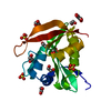
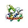
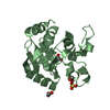
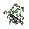
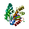
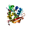
 Pseudomonas aeruginosa (bacteria)
 X-RAY DIFFRACTION /
  Authors
 Citation
 Structure visualization
 Downloads & links
 Other downloads
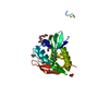


 PDBj
PDBj

 Assembly

 Mass: 96.063 Da / Num. of mol.: 2 / Source method: obtained synthetically / Formula: SO4
 Mass: 35.453 Da / Num. of mol.: 4 / Source method: obtained synthetically / Formula: Cl
 Mass: 238.305 Da / Num. of mol.: 1 / Source method: obtained synthetically / Formula: C8H18N2O4S / Comment: pH buffer*YM
 Mass: 18.015 Da / Num. of mol.: 312 / Source method: isolated from a natural source / Formula: H2O
 Sample preparation
 / Beamline: 19-ID / Wavelength: 0.97932 Å
 Processing