[English] 日本語
 Yorodumi
- PDB-4m32: Crystal structure of gated-pore mutant D138N of second DNA-Bindin... -
+ Open data
- Basic information
| Entry | Database: PDB / ID: 4m32 | ||||||
|---|---|---|---|---|---|---|---|
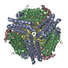
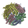
| Title | Crystal structure of gated-pore mutant D138N of second DNA-Binding protein under starvation from Mycobacterium smegmatis | ||||||
 Components Components | Putative starvation-induced DNA protecting protein/Ferritin and Dps | ||||||
 Keywords Keywords | DNA BINDING PROTEIN / ferritin-like fold / ferroxidation / Iron | ||||||
| Function / homology |  Function and homology information Function and homology informationoxidoreductase activity, acting on metal ions / ferric iron binding / DNA binding Similarity search - Function | ||||||
| Biological species |  Mycobacterium smegmatis (bacteria) Mycobacterium smegmatis (bacteria) | ||||||
| Method |  X-RAY DIFFRACTION / Direct refinement against model PDB / Resolution: 1.86 Å X-RAY DIFFRACTION / Direct refinement against model PDB / Resolution: 1.86 Å | ||||||
 Authors Authors | Williams, S.M. / Chandran, A.V. / Vijayabaskar, M.S. / Roy, S. / Balaram, H. / Vishveshwara, S. / Vijayan, M. / Chatterji, D. | ||||||
 Citation Citation |  Journal: J.Biol.Chem. / Year: 2014 Journal: J.Biol.Chem. / Year: 2014Title: A histidine aspartate ionic lock gates the iron passage in miniferritins from Mycobacterium smegmatis Authors: Williams, S.M. / Chandran, A.V. / Vijayabaskar, M.S. / Roy, S. / Balaram, H. / Vishveshwara, S. / Vijayan, M. / Chatterji, D. | ||||||
| History |
|
- Structure visualization
| Structure viewer | Molecule:  Molmil Molmil Jmol/JSmol Jmol/JSmol |
|---|
- Downloads & links
- Download
| PDBx/mmCIF format |  4m32.cif.gz 4m32.cif.gz | 141.9 KB | Display |  PDBx/mmCIF format PDBx/mmCIF format |
|---|---|---|---|---|
| PDB format |  pdb4m32.ent.gz pdb4m32.ent.gz | 111.7 KB | Display |  PDB format PDB format |
| PDBx/mmJSON format |  4m32.json.gz 4m32.json.gz | Tree view |  PDBx/mmJSON format PDBx/mmJSON format | |
| Others |  Other downloads Other downloads |
-Validation report
| Summary document |  4m32_validation.pdf.gz 4m32_validation.pdf.gz | 447.7 KB | Display |  wwPDB validaton report wwPDB validaton report |
|---|---|---|---|---|
| Full document |  4m32_full_validation.pdf.gz 4m32_full_validation.pdf.gz | 450.8 KB | Display | |
| Data in XML |  4m32_validation.xml.gz 4m32_validation.xml.gz | 27.1 KB | Display | |
| Data in CIF |  4m32_validation.cif.gz 4m32_validation.cif.gz | 39.5 KB | Display | |
| Arichive directory |  https://data.pdbj.org/pub/pdb/validation_reports/m3/4m32 https://data.pdbj.org/pub/pdb/validation_reports/m3/4m32 ftp://data.pdbj.org/pub/pdb/validation_reports/m3/4m32 ftp://data.pdbj.org/pub/pdb/validation_reports/m3/4m32 | HTTPS FTP |
-Related structure data
| Related structure data | 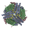 4m33C  4m34C  4m35C 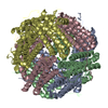 2z90S C: citing same article ( S: Starting model for refinement |
|---|---|
| Similar structure data |
- Links
Links
- Assembly
| Deposited unit | 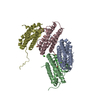
| |||||||||||||||||||||
|---|---|---|---|---|---|---|---|---|---|---|---|---|---|---|---|---|---|---|---|---|---|---|
| 1 | 
| |||||||||||||||||||||
| Unit cell |
| |||||||||||||||||||||
| Components on special symmetry positions |
|
- Components
| #1: Protein | Mass: 18511.699 Da / Num. of mol.: 4 / Mutation: D138N Source method: isolated from a genetically manipulated source Source: (gene. exp.)  Mycobacterium smegmatis (bacteria) / Strain: ATCC 700084 / mc(2)155 / Gene: DPS2, MSMEG_3242, MSMEI_3159 / Plasmid: pET21b / Production host: Mycobacterium smegmatis (bacteria) / Strain: ATCC 700084 / mc(2)155 / Gene: DPS2, MSMEG_3242, MSMEI_3159 / Plasmid: pET21b / Production host:  #2: Chemical | #3: Chemical | #4: Chemical | ChemComp-FE2 / #5: Water | ChemComp-HOH / | |
|---|
-Experimental details
-Experiment
| Experiment | Method:  X-RAY DIFFRACTION / Number of used crystals: 1 X-RAY DIFFRACTION / Number of used crystals: 1 |
|---|
- Sample preparation
| Crystal | Density Matthews: 2.2 Å3/Da / Density % sol: 44.06 % Description: THE ENTRY CONTAINS FRIEDEL PAIRS IN F_PLUS/MINUS COLUMNS. |
|---|---|
| Crystal grow | Temperature: 298 K / Method: microbatch under oil / pH: 6.5 Details: 150mM MgCl2, 0.1M sodium cacodylate, 20% PEG3350, pH 6.5, Microbatch under oil, temperature 298K |
-Data collection
| Diffraction | Mean temperature: 100 K |
|---|---|
| Diffraction source | Source:  ROTATING ANODE / Type: BRUKER AXS MICROSTAR / Wavelength: 1.54179 Å ROTATING ANODE / Type: BRUKER AXS MICROSTAR / Wavelength: 1.54179 Å |
| Detector | Type: MAR scanner 345 mm plate / Detector: IMAGE PLATE / Date: Aug 27, 2011 / Details: mirrors |
| Radiation | Monochromator: Osmic / Protocol: SINGLE WAVELENGTH / Monochromatic (M) / Laue (L): M / Scattering type: x-ray |
| Radiation wavelength | Wavelength: 1.54179 Å / Relative weight: 1 |
| Reflection | Resolution: 1.85→43.57 Å / Num. obs: 55022 / Redundancy: 4.6 % / Biso Wilson estimate: 19.5 Å2 / Rmerge(I) obs: 0.064 / Net I/σ(I): 15.3 |
| Reflection shell | Resolution: 1.85→1.95 Å / Redundancy: 5 % / Rmerge(I) obs: 0.232 / Mean I/σ(I) obs: 5.6 |
- Processing
| Software |
| |||||||||||||||||||||||||||||||||||||||||||||||||||||||||||||||||
|---|---|---|---|---|---|---|---|---|---|---|---|---|---|---|---|---|---|---|---|---|---|---|---|---|---|---|---|---|---|---|---|---|---|---|---|---|---|---|---|---|---|---|---|---|---|---|---|---|---|---|---|---|---|---|---|---|---|---|---|---|---|---|---|---|---|---|
| Refinement | Method to determine structure: Direct refinement against model PDB Starting model: 2z90 Resolution: 1.86→30 Å / Cor.coef. Fo:Fc: 0.951 / Cor.coef. Fo:Fc free: 0.939 / SU B: 2.557 / SU ML: 0.079 / Cross valid method: THROUGHOUT / ESU R: 0.145 / ESU R Free: 0.127 / Stereochemistry target values: MAXIMUM LIKELIHOOD Details: SF FILE CONTAINS FRIEDEL PAIRS UNDER I/F_MINUS AND I/F_PLUS COLUMNS.
| |||||||||||||||||||||||||||||||||||||||||||||||||||||||||||||||||
| Solvent computation | Ion probe radii: 0.8 Å / Shrinkage radii: 0.8 Å / VDW probe radii: 1.4 Å / Solvent model: MASK | |||||||||||||||||||||||||||||||||||||||||||||||||||||||||||||||||
| Displacement parameters | Biso mean: 15.327 Å2
| |||||||||||||||||||||||||||||||||||||||||||||||||||||||||||||||||
| Refinement step | Cycle: LAST / Resolution: 1.86→30 Å
| |||||||||||||||||||||||||||||||||||||||||||||||||||||||||||||||||
| Refine LS restraints |
| |||||||||||||||||||||||||||||||||||||||||||||||||||||||||||||||||
| LS refinement shell | Resolution: 1.86→1.91 Å / Total num. of bins used: 20
|
 Movie
Movie Controller








 PDBj
PDBj


