- PDB-5ww8: Crystal structure of the second DNA-Binding protein under starvat... -
+
Open data
ID or keywords:
Loading...
-
Basic information
Entry
Database: PDB / ID: 5ww8
Title
Crystal structure of the second DNA-Binding protein under starvation from Mycobacterium smegmatis soaked with iron in the ratio of 480 iron atoms per dodecamer
Components
Putative starvation-induced DNA protecting protein/Ferritin and Dps
Keywords
DNA BINDING PROTEIN / ferritin-like fold / DNA binding / ferroxidation / OXIDOREDUCTASE
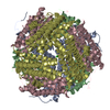
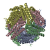
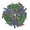
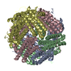
A: Putative starvation-induced DNA protecting protein/Ferritin and Dps B: Putative starvation-induced DNA protecting protein/Ferritin and Dps C: Putative starvation-induced DNA protecting protein/Ferritin and Dps D: Putative starvation-induced DNA protecting protein/Ferritin and Dps hetero molecules
A: Putative starvation-induced DNA protecting protein/Ferritin and Dps B: Putative starvation-induced DNA protecting protein/Ferritin and Dps C: Putative starvation-induced DNA protecting protein/Ferritin and Dps D: Putative starvation-induced DNA protecting protein/Ferritin and Dps hetero molecules
A: Putative starvation-induced DNA protecting protein/Ferritin and Dps B: Putative starvation-induced DNA protecting protein/Ferritin and Dps C: Putative starvation-induced DNA protecting protein/Ferritin and Dps D: Putative starvation-induced DNA protecting protein/Ferritin and Dps hetero molecules
A: Putative starvation-induced DNA protecting protein/Ferritin and Dps B: Putative starvation-induced DNA protecting protein/Ferritin and Dps C: Putative starvation-induced DNA protecting protein/Ferritin and Dps D: Putative starvation-induced DNA protecting protein/Ferritin and Dps hetero molecules
Mass: 18.015 Da / Num. of mol.: 246 / Source method: isolated from a natural source / Formula: H2O
-
Experimental details
-
Experiment
Experiment
Method: X-RAY DIFFRACTION / Number of used crystals: 1
-
Sample preparation
Crystal
Density Matthews: 2.3 Å3/Da / Density % sol: 47.21 %
Crystal grow
Temperature: 298 K / Method: microbatch / pH: 6 Details: 100 mM sodium cacodylate, 150 mM MgCl2, 20% PEG3000
-
Data collection
Diffraction
Mean temperature: 100 K
Diffraction source
Source: ROTATING ANODE / Type: BRUKER AXS MICROSTAR / Wavelength: 1.54179 Å
Detector
Type: MAR scanner 345 mm plate / Detector: IMAGE PLATE / Date: Feb 24, 2012
Radiation
Protocol: SINGLE WAVELENGTH / Monochromatic (M) / Laue (L): M / Scattering type: x-ray
Radiation wavelength
Wavelength: 1.54179 Å / Relative weight: 1
Reflection
Resolution: 2.03→30 Å / Num. obs: 42006 / % possible obs: 98.2 % / Redundancy: 13.7 % / Net I/σ(I): 19
Reflection shell
Resolution: 2.03→2.14 Å / Redundancy: 13.9 % / Mean I/σ(I) obs: 3.4 / % possible all: 87.9
-
Processing
Software
Name
Version
Classification
REFMAC
5.8.0103
refinement
MOSFLM
datareduction
SCALA
datascaling
REFMAC
phasing
Refinement
Resolution: 2.04→30 Å / Cor.coef. Fo:Fc: 0.95 / Cor.coef. Fo:Fc free: 0.929 / SU B: 4.04 / SU ML: 0.108 / Cross valid method: THROUGHOUT / ESU R: 0.195 / ESU R Free: 0.159 / Details: HYDROGENS HAVE BEEN USED IF PRESENT IN THE INPUT
Rfactor
Num. reflection
% reflection
Selection details
Rfree
0.21043
2120
5 %
RANDOM
Rwork
0.17748
-
-
-
obs
0.17909
39886
99.82 %
-
Solvent computation
Ion probe radii: 0.8 Å / Shrinkage radii: 0.8 Å / VDW probe radii: 1.2 Å
 Movie
Movie Controller

 Yorodumi
 Open data
 Basic information
 Components
 Keywords
 Function and homology information
 Mycobacterium smegmatis str. MC2 155 (bacteria)
 X-RAY DIFFRACTION / Resolution: 2.04 Å
  Authors
 Citation
 Structure visualization
 Downloads & links
 Other downloads




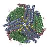
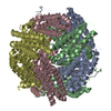
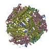
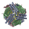
 PDBj
PDBj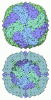
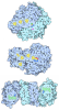

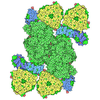
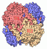

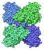
 Assembly
 Escherichia coli (E. coli) / References: UniProt: A0QXB7




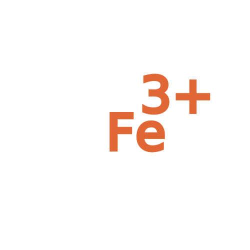 Mass: 55.845 Da / Num. of mol.: 4 / Source method: obtained synthetically / Formula: Fe
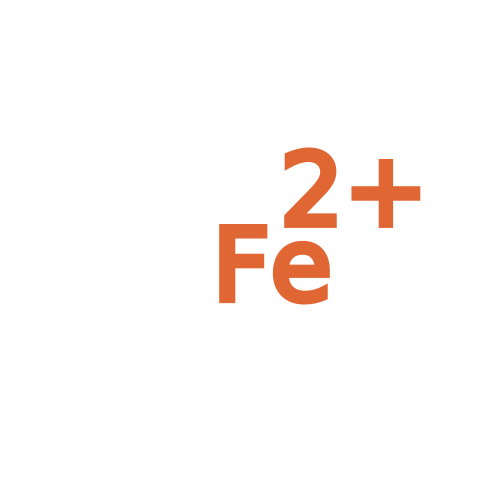 Mass: 55.845 Da / Num. of mol.: 4 / Source method: obtained synthetically / Formula: Fe
 Mass: 24.305 Da / Num. of mol.: 2 / Source method: obtained synthetically / Formula: Mg
 Mass: 35.453 Da / Num. of mol.: 2 / Source method: obtained synthetically / Formula: Cl
 Sample preparation
 Processing