 Movie
Movie Controller

[English] 日本語
 Yorodumi
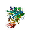
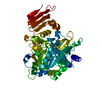
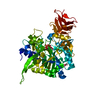
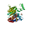
- PDB-4m2a: Crystal structure of the udp-glucose pyrophosphorylase from Leish... -
+ Open data

- Basic information
| Entry | Database: PDB / ID: 4m2a | ||||||
|---|---|---|---|---|---|---|---|
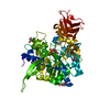
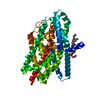
| Title | Crystal structure of the udp-glucose pyrophosphorylase from Leishmania major in the post-reactive state | ||||||
 Components Components | UDP-glucose pyrophosphorylase | ||||||
 Keywords Keywords | TRANSFERASE / ROSSMANN-LIKE ALPHA/BETA/ALPHA SANDWICH FOLD / NUCLEOTIDYLTRANSFERASE | ||||||
| Function / homology |  Function and homology information Function and homology informationUTP-glucose-1-phosphate uridylyltransferase / UTP:glucose-1-phosphate uridylyltransferase activity / UDP-alpha-D-glucose metabolic process / nuclear lumen / ciliary plasm / glycogen metabolic process / cytosol / cytoplasm Similarity search - Function | ||||||
| Biological species |  Leishmania major (eukaryote) Leishmania major (eukaryote) | ||||||
| Method |  X-RAY DIFFRACTION / SYNCHROTRON / MOLECULAR REPLACEMENT / Resolution: 1.66 Å X-RAY DIFFRACTION / SYNCHROTRON / MOLECULAR REPLACEMENT / Resolution: 1.66 Å | ||||||
 Authors Authors | Fuehring, J. / Routier, F.H. / Lamerz, A.-C. / Baruch, P. / Gerardy-Schahn, R. / Fedorov, R. | ||||||
 Citation Citation | Journal: ACS CATALYSIS / Year: 2013 Title: Catalytic Mechanism and Allosteric Regulation of Udp-Glucose Pyrophosphorylase from Leishmania Major Authors: Fuehring, J. / Routier, F.H. / Lamerz, A.-C. / Baruch, P. / Gerardy-Schahn, R. / Fedorov, R. | ||||||
| History |
|
- Structure visualization
| Structure viewer | Molecule: MolmilJmol/JSmol |
|---|
- Downloads & links
-Download
| PDBx/mmCIF format | 4m2a.cif.gz | 125.2 KB | Display | PDBx/mmCIF format |
|---|---|---|---|---|
| PDB format | pdb4m2a.ent.gz | 94.1 KB | Display | PDB format |
| PDBx/mmJSON format | 4m2a.json.gz | Tree view | PDBx/mmJSON format | |
| Others |  Other downloads Other downloads |
-Validation report
| Arichive directory | https://data.pdbj.org/pub/pdb/validation_reports/m2/4m2aftp://data.pdbj.org/pub/pdb/validation_reports/m2/4m2a | HTTPS FTP |
|---|
-Related structure data
| Related structure data |  4m2bC 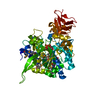 2oegS S: Starting model for refinement C: citing same article ( |
|---|---|
| Similar structure data |


-Links
 PDBj
PDBj

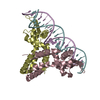
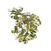
- Assembly
| Deposited unit | 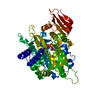
| |||||||||
|---|---|---|---|---|---|---|---|---|---|---|
| 1 |
| |||||||||
| Unit cell |
| |||||||||
| Components on special symmetry positions |
| |||||||||
| Details | BIOMOLECULE: 1 SEE REMARK 350 FOR THE AUTHOR PROVIDED AND/OR PROGRAM GENERATED ASSEMBLY INFORMATION FOR THE STRUCTURE IN THIS ENTRY. THE REMARK MAY ALSO PROVIDE INFORMATION ON BURIED SURFACE AREA. COORDINATES FOR A COMPLETE MULTIMER REPRESENTING THE KNOWN BIOLOGICALLY SIGNIFICANT OLIGOMERIZATION STATE OF THE MOLECULE CAN BE GENERATED BY APPLYING BIOMT TRANSFORMATIONS GIVEN BELOW. BOTH NON-CRYSTALLOGRAPHIC AND CRYSTALLOGRAPHIC OPERATIONS ARE GIVEN. BIOMOLECULE: 1 AUTHOR DETERMINED BIOLOGICAL UNIT: MONOMERIC SOFTWARE DETERMINED QUATERNARY STRUCTURE: MONOMERIC SOFTWARE USED: PISA APPLY THE FOLLOWING TO CHAINS: A BIOMT1 1 1.000000 0.000000 0.000000 0.00000 BIOMT2 1 0.000000 1.000000 0.000000 0.00000 BIOMT3 1 0.000000 0.000000 1.000000 0.00000 |
-Components
| #1: Protein | Mass: 56054.770 Da / Num. of mol.: 1 Source method: isolated from a genetically manipulated source Source: (gene. exp.) Leishmania major (eukaryote) / Gene: LMJF_18_0990, UGP / Production host:  References: UniProt: Q4QDU3, UTP-glucose-1-phosphate uridylyltransferase |
|---|---|
| #2: Chemical | ChemComp-UPG /   Mass: 566.302 Da / Num. of mol.: 1 / Source method: obtained synthetically / Formula: C15H24N2O17P2 Mass: 566.302 Da / Num. of mol.: 1 / Source method: obtained synthetically / Formula: C15H24N2O17P2 |
| #3: Chemical | ChemComp-MG /   Mass: 24.305 Da / Num. of mol.: 1 / Source method: obtained synthetically / Formula: Mg Mass: 24.305 Da / Num. of mol.: 1 / Source method: obtained synthetically / Formula: Mg |
| #4: Chemical | ChemComp-SO4 /   Mass: 96.063 Da / Num. of mol.: 1 / Source method: obtained synthetically / Formula: SO4 Mass: 96.063 Da / Num. of mol.: 1 / Source method: obtained synthetically / Formula: SO4 |
| #5: Water | ChemComp-HOH /  Mass: 18.015 Da / Num. of mol.: 665 / Source method: isolated from a natural source / Formula: H2O Mass: 18.015 Da / Num. of mol.: 665 / Source method: isolated from a natural source / Formula: H2O |
-Experimental details
-Experiment
| Experiment | Method: X-RAY DIFFRACTION / Number of used crystals: 1 |
|---|
- Sample preparation
| Crystal | Density Matthews: 2.16 Å3/Da / Density % sol: 43.15 % |
|---|---|
| Crystal grow | Method: vapor diffusion, hanging drop / pH: 6 Details: 0.1M BIS-TRIS, 22% W/V PEG-MME- 2000, 20MM (NH4)2SO4, pH 6.0, VAPOR DIFFUSION, HANGING DROP |
-Data collection
| Diffraction | Mean temperature: 100 K | |||||||||
|---|---|---|---|---|---|---|---|---|---|---|
| Diffraction source | Source: SYNCHROTRON / Site: EMBL/DESY, HAMBURG  / Beamline: X13 / Wavelength: 0.806 / Wavelength: 0.8055 Å / Beamline: X13 / Wavelength: 0.806 / Wavelength: 0.8055 Å | |||||||||
| Detector | Type: MAR CCD 165 mm / Detector: CCD / Date: Oct 31, 2007 | |||||||||
| Radiation | Monochromator: THE TRIANGULAR SHAPED GE (OR SI) MONOCHROMATOR CUT AT 7 FOR COMPRESSION OF THE BEAM IS 180 MM LONG, 1 MM THICK AND 40 MM IN HEIGHT AT THE BASE. Protocol: SINGLE WAVELENGTH / Monochromatic (M) / Laue (L): M / Scattering type: x-ray | |||||||||
| Radiation wavelength |
| |||||||||
| Reflection | Resolution: 1.66→29.6 Å / Num. all: 57313 / Num. obs: 57313 / % possible obs: 99.7 % / Observed criterion σ(F): 0 / Observed criterion σ(I): 2 / Redundancy: 14.93 % / Biso Wilson estimate: 26.4 Å2 / Rmerge(I) obs: 0.068 / Rsym value: 0.023 / Net I/σ(I): 28.78 |
- Processing
| Software |
| ||||||||||||||||||||
|---|---|---|---|---|---|---|---|---|---|---|---|---|---|---|---|---|---|---|---|---|---|
| Refinement | Method to determine structure: MOLECULAR REPLACEMENT Starting model: PDB ENTRY 2OEG Resolution: 1.66→29.6 Å / Cross valid method: THROUGHOUT / σ(F): 0 / Stereochemistry target values: Engh & Huber
| ||||||||||||||||||||
| Refinement step | Cycle: LAST / Resolution: 1.66→29.6 Å
|
