 Movie
Movie Controller

[English] 日本語
 Yorodumi
- PDB-4m1r: Structure of a novel cellulase 5 from a sugarcane soil metagenomi... -
+ Open data

- Basic information
| Entry | Database: PDB / ID: 4m1r | ||||||
|---|---|---|---|---|---|---|---|
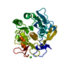
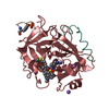
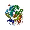
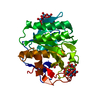
| Title | Structure of a novel cellulase 5 from a sugarcane soil metagenomic library | ||||||
 Components Components | Cellulase 5 | ||||||
 Keywords Keywords | HYDROLASE / TIM barrel / cellulase | ||||||
| Function / homology | Glycosidases / TIM Barrel / Alpha-Beta Barrel / Alpha Beta Function and homology information Function and homology information | ||||||
| Biological species | soil metagenome (others) | ||||||
| Method |  X-RAY DIFFRACTION / SYNCHROTRON / MOLECULAR REPLACEMENT / Resolution: 1.8 Å X-RAY DIFFRACTION / SYNCHROTRON / MOLECULAR REPLACEMENT / Resolution: 1.8 Å | ||||||
 Authors Authors | Paiva, J.H. / Alvarez, T.M. / Cairo, J.P. / Paixao, D.A. / Almeida, R.A. / Tonoli, C.C.C. / Ruiz, D.M. / Ruller, R. / Santos, C.R. / Squina, F.M. / Murakami, M.T. | ||||||
 Citation Citation | Journal: Plos One / Year: 2013 Title: Structure and function of a novel cellulase 5 from sugarcane soil metagenome. Authors: Alvarez, T.M. / Paiva, J.H. / Ruiz, D.M. / Cairo, J.P. / Pereira, I.O. / Paixao, D.A. / de Almeida, R.F. / Tonoli, C.C. / Ruller, R. / Santos, C.R. / Squina, F.M. / Murakami, M.T. | ||||||
| History |
|
- Structure visualization
| Structure viewer | Molecule: MolmilJmol/JSmol |
|---|
- Downloads & links
-Download
| PDBx/mmCIF format | 4m1r.cif.gz | 230.7 KB | Display | PDBx/mmCIF format |
|---|---|---|---|---|
| PDB format | pdb4m1r.ent.gz | 187.5 KB | Display | PDB format |
| PDBx/mmJSON format | 4m1r.json.gz | Tree view | PDBx/mmJSON format | |
| Others |  Other downloads Other downloads |
-Validation report
| Arichive directory | https://data.pdbj.org/pub/pdb/validation_reports/m1/4m1rftp://data.pdbj.org/pub/pdb/validation_reports/m1/4m1r | HTTPS FTP |
|---|
-Related structure data
| Similar structure data |
|---|


-Links
 PDBj
PDBj- Assembly
| Deposited unit | 
| ||||||||
|---|---|---|---|---|---|---|---|---|---|
| 1 | 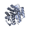
| ||||||||
| 2 | 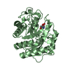
| ||||||||
| Unit cell |
|
-Components
| #1: Protein | Mass: 31772.094 Da / Num. of mol.: 2 Source method: isolated from a genetically manipulated source Source: (gene. exp.) soil metagenome (others) / Production host:  #2: Chemical |   Mass: 122.143 Da / Num. of mol.: 2 / Source method: obtained synthetically / Formula: C4H12NO3 / Comment: pH buffer*YM Mass: 122.143 Da / Num. of mol.: 2 / Source method: obtained synthetically / Formula: C4H12NO3 / Comment: pH buffer*YM#3: Water | ChemComp-HOH / |  Mass: 18.015 Da / Num. of mol.: 389 / Source method: isolated from a natural source / Formula: H2O Mass: 18.015 Da / Num. of mol.: 389 / Source method: isolated from a natural source / Formula: H2O |
|---|
-Experimental details
-Experiment
| Experiment | Method: X-RAY DIFFRACTION / Number of used crystals: 1 |
|---|
- Sample preparation
| Crystal | Density Matthews: 1.9 Å3/Da / Density % sol: 35.28 % |
|---|---|
| Crystal grow | Temperature: 291 K / Method: vapor diffusion, sitting drop / pH: 8.5 Details: 35% (v/v) isopropanol, 30% (w/v) PEG3350 and 100 mM Tris, pH 8.5, VAPOR DIFFUSION, SITTING DROP, temperature 291K |
-Data collection
| Diffraction | Mean temperature: 100 K |
|---|---|
| Diffraction source | Source: SYNCHROTRON / Site: LNLS  / Beamline: W01B-MX2 / Wavelength: 1.458 Å / Beamline: W01B-MX2 / Wavelength: 1.458 Å |
| Detector | Type: MARMOSAIC 225 mm CCD / Detector: CCD / Date: Dec 10, 2012 |
| Radiation | Monochromator: Double-crystal Si 111 / Protocol: SINGLE WAVELENGTH / Monochromatic (M) / Laue (L): M / Scattering type: x-ray |
| Radiation wavelength | Wavelength: 1.458 Å / Relative weight: 1 |
| Reflection | Resolution: 1.8→50 Å / Num. all: 41174 / Num. obs: 41174 / % possible obs: 93.5 % / Observed criterion σ(F): 1 / Observed criterion σ(I): 1 |
- Processing
| Software |
| |||||||||||||||||||||||||||||||||||||||||||||||||||||||||||||||||||||||||||
|---|---|---|---|---|---|---|---|---|---|---|---|---|---|---|---|---|---|---|---|---|---|---|---|---|---|---|---|---|---|---|---|---|---|---|---|---|---|---|---|---|---|---|---|---|---|---|---|---|---|---|---|---|---|---|---|---|---|---|---|---|---|---|---|---|---|---|---|---|---|---|---|---|---|---|---|---|
| Refinement | Method to determine structure: MOLECULAR REPLACEMENT / Resolution: 1.8→28.55 Å / Cor.coef. Fo:Fc: 0.973 / Cor.coef. Fo:Fc free: 0.958 / SU B: 4.703 / SU ML: 0.067 / Cross valid method: THROUGHOUT / ESU R: 0.123 / ESU R Free: 0.115 / Stereochemistry target values: MAXIMUM LIKELIHOOD
| |||||||||||||||||||||||||||||||||||||||||||||||||||||||||||||||||||||||||||
| Solvent computation | Ion probe radii: 0.8 Å / Shrinkage radii: 0.8 Å / VDW probe radii: 1.4 Å / Solvent model: MASK | |||||||||||||||||||||||||||||||||||||||||||||||||||||||||||||||||||||||||||
| Displacement parameters | Biso mean: 15.097 Å2
| |||||||||||||||||||||||||||||||||||||||||||||||||||||||||||||||||||||||||||
| Refinement step | Cycle: LAST / Resolution: 1.8→28.55 Å
| |||||||||||||||||||||||||||||||||||||||||||||||||||||||||||||||||||||||||||
| Refine LS restraints |
| |||||||||||||||||||||||||||||||||||||||||||||||||||||||||||||||||||||||||||
| LS refinement shell | Resolution: 1.799→1.846 Å / Total num. of bins used: 20
| |||||||||||||||||||||||||||||||||||||||||||||||||||||||||||||||||||||||||||
| Refinement TLS params. | Method: refined / Refine-ID: X-RAY DIFFRACTION
| |||||||||||||||||||||||||||||||||||||||||||||||||||||||||||||||||||||||||||
| Refinement TLS group |
|
