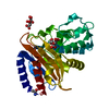
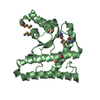
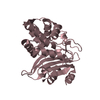
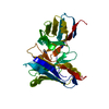
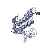
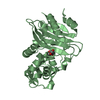
[English] 日本語
Yorodumi
- PDB-4lrn: Ontogeny of recognition specificity and functionality for the ant... -

+
Open data


ID or keywords:

Loading...

-
Basic information

Entry
Database: PDB / ID: 4lrn
TitleOntogeny of recognition specificity and functionality for the anti-HIV antibody 4E10
Components
  • GEP 1 heavy chain
  • GEP 1 light chain
KeywordsIMMUNE SYSTEM / germline / HIV / immunoglobulin / Fv portion of Ab / 4E10 epitope
Function / homology
Function and homology information


pentameric IgM immunoglobulin complex / monomeric IgA immunoglobulin complex / secretory IgA immunoglobulin complex / glomerular filtration / CD22 mediated BCR regulation / Fc epsilon receptor (FCERI) signaling / Classical antibody-mediated complement activation / Initial triggering of complement / FCGR activation / Role of phospholipids in phagocytosis ...pentameric IgM immunoglobulin complex / monomeric IgA immunoglobulin complex / secretory IgA immunoglobulin complex / glomerular filtration / CD22 mediated BCR regulation / Fc epsilon receptor (FCERI) signaling / Classical antibody-mediated complement activation / Initial triggering of complement / FCGR activation / Role of phospholipids in phagocytosis / Role of LAT2/NTAL/LAB on calcium mobilization / Scavenging of heme from plasma / FCERI mediated Ca+2 mobilization / FCGR3A-mediated IL10 synthesis / Antigen activates B Cell Receptor (BCR) leading to generation of second messengers / Regulation of Complement cascade / Cell surface interactions at the vascular wall / FCERI mediated MAPK activation / FCGR3A-mediated phagocytosis / antigen binding / Regulation of actin dynamics for phagocytic cup formation / FCERI mediated NF-kB activation / Immunoregulatory interactions between a Lymphoid and a non-Lymphoid cell / antibacterial humoral response / adaptive immune response / Potential therapeutics for SARS / blood microparticle / immune response / extracellular space / extracellular exosome / extracellular region / plasma membrane
Similarity search - Function
Immunoglobulin V-Type / Immunoglobulin V-set domain / Immunoglobulin V-set domain / Immunoglobulin subtype / Immunoglobulin / Ig-like domain profile. / Immunoglobulin-like domain / Immunoglobulin-like domain superfamily / Immunoglobulins / Immunoglobulin-like fold ...Immunoglobulin V-Type / Immunoglobulin V-set domain / Immunoglobulin V-set domain / Immunoglobulin subtype / Immunoglobulin / Ig-like domain profile. / Immunoglobulin-like domain / Immunoglobulin-like domain superfamily / Immunoglobulins / Immunoglobulin-like fold / Immunoglobulin-like / Sandwich / Mainly Beta
Similarity search - Domain/homology
Immunoglobulin kappa variable 3-20
Similarity search - Component
Biological speciesHomo sapiens (human)
MethodX-RAY DIFFRACTION / SYNCHROTRON / MOLECULAR REPLACEMENT / Resolution: 1.89 Å
AuthorsFinton, K.A.K.
CitationJournal: Plos Pathog. / Year: 2014
Title: Ontogeny of Recognition Specificity and Functionality for the Broadly Neutralizing Anti-HIV Antibody 4E10.
Authors: Finton, K.A. / Friend, D. / Jaffe, J. / Gewe, M. / Holmes, M.A. / Larman, H.B. / Stuart, A. / Larimore, K. / Greenberg, P.D. / Elledge, S.J. / Stamatatos, L. / Strong, R.K.
History
DepositionJul 20, 2013Deposition site: RCSB / Processing site: RCSB
Revision 1.0Oct 8, 2014Provider: repository / Type: Initial release

-
Structure visualization

Structure viewerMolecule:
MolmilJmol/JSmol

Downloads & links

-
Assembly

Deposited unit
H: GEP 1 heavy chain
L: GEP 1 light chain


Theoretical massNumber of molelcules
Total (without water)25,5022
Polymers25,5022
Non-polymers00
Water1,910106
1


  • Idetical with deposited unit
  • defined by author&software
TypeNameSymmetry operationNumber
identity operation1_555x,y,z1
Buried area1380 Å2
ΔGint-11 kcal/mol
Surface area10890 Å2
MethodPISA
Unit cell
Length a, b, c (Å)35.494, 48.733, 110.584
Angle α, β, γ (deg.)90.00, 90.00, 90.00
Int Tables number19
Space group name H-MP212121

-
Components

#1: Antibody GEP 1 heavy chain


Mass: 13512.175 Da / Num. of mol.: 1
Source method: isolated from a genetically manipulated source
Source: (gene. exp.) Homo sapiens (human) / Production host: Escherichia coli (E. coli)
#2: Antibody GEP 1 light chain


Mass: 11989.346 Da / Num. of mol.: 1
Source method: isolated from a genetically manipulated source
Source: (gene. exp.) Homo sapiens (human) / Production host: Escherichia coli (E. coli) / References: UniProt: P01619*PLUS
#3: Water ChemComp-HOH / water


Mass: 18.015 Da / Num. of mol.: 106 / Source method: isolated from a natural source / Formula: H2O

-
Experimental details

-
Experiment

ExperimentMethod: X-RAY DIFFRACTION / Number of used crystals: 1

-
Sample preparation

CrystalDensity Matthews: 1.88 Å3/Da / Density % sol: 34.41 %
Crystal growTemperature: 298 K / Method: vapor diffusion, hanging drop / pH: 8.5
Details: 0.2M NaCl, 0.1 M Tris pH 8.5, 25%, w/v PEG 3350, VAPOR DIFFUSION, HANGING DROP, temperature 298K

-
Data collection

DiffractionMean temperature: 100 K
Diffraction sourceSource: SYNCHROTRON / Site: ALS / Beamline: 5.0.1 / Wavelength: 1 Å
DetectorType: ADSC QUANTUM 315r / Detector: CCD / Date: Aug 24, 2012
RadiationMonochromator: Si(220) Asymmetric cut single crystal / Protocol: SINGLE WAVELENGTH / Monochromatic (M) / Laue (L): M / Scattering type: x-ray
Radiation wavelengthWavelength: 1 Å / Relative weight: 1
ReflectionResolution: 1.89→50 Å / Num. all: 15973 / Num. obs: 15973 / % possible obs: 100 %
Reflection shellResolution: 1.89→1.93 Å / % possible all: 100

-
Processing

Software
NameVersionClassification
ADSCQuantumdata collection
PHASERphasing
REFMAC5.7.0029refinement
HKL-2000data reduction
HKL-2000data scaling
RefinementMethod to determine structure: MOLECULAR REPLACEMENT / Resolution: 1.89→48.78 Å / Cor.coef. Fo:Fc: 0.953 / Cor.coef. Fo:Fc free: 0.927 / SU B: 7.414 / SU ML: 0.108 / Cross valid method: THROUGHOUT / ESU R: 0.167 / ESU R Free: 0.153 / Stereochemistry target values: MAXIMUM LIKELIHOOD / Details: HYDROGENS HAVE BEEN ADDED IN THE RIDING POSITIONS
RfactorNum. reflection% reflectionSelection details
Rfree0.23283 796 5 %RANDOM
Rwork0.18739 ---
obs0.18963 15126 99.66 %-
all-15973 --
Solvent computationIon probe radii: 0.8 Å / Shrinkage radii: 0.8 Å / VDW probe radii: 1.2 Å / Solvent model: MASK
Displacement parametersBiso mean: 24.711 Å2
Baniso -1Baniso -2Baniso -3
1--2.91 Å20 Å20 Å2
2--4.57 Å20 Å2
3----1.66 Å2
Refinement stepCycle: LAST / Resolution: 1.89→48.78 Å
ProteinNucleic acidLigandSolventTotal
Num. atoms1630 0 0 106 1736
Refine LS restraints
Refine-IDTypeDev idealDev ideal targetNumber
X-RAY DIFFRACTIONr_bond_refined_d0.0040.021685
X-RAY DIFFRACTIONr_bond_other_d00.021525
X-RAY DIFFRACTIONr_angle_refined_deg0.7651.9582299
X-RAY DIFFRACTIONr_angle_other_deg0.5183.0023510
X-RAY DIFFRACTIONr_dihedral_angle_1_deg6.885230
X-RAY DIFFRACTIONr_dihedral_angle_2_deg28.25723.63655
X-RAY DIFFRACTIONr_dihedral_angle_3_deg13.3915242
X-RAY DIFFRACTIONr_dihedral_angle_4_deg14.319156
X-RAY DIFFRACTIONr_chiral_restr0.0610.2260
X-RAY DIFFRACTIONr_gen_planes_refined0.0010.0211934
X-RAY DIFFRACTIONr_gen_planes_other00.02372
X-RAY DIFFRACTIONr_nbd_refined
X-RAY DIFFRACTIONr_nbd_other
X-RAY DIFFRACTIONr_nbtor_refined
X-RAY DIFFRACTIONr_nbtor_other
X-RAY DIFFRACTIONr_xyhbond_nbd_refined
X-RAY DIFFRACTIONr_xyhbond_nbd_other
X-RAY DIFFRACTIONr_metal_ion_refined
X-RAY DIFFRACTIONr_metal_ion_other
X-RAY DIFFRACTIONr_symmetry_vdw_refined
X-RAY DIFFRACTIONr_symmetry_vdw_other
X-RAY DIFFRACTIONr_symmetry_hbond_refined
X-RAY DIFFRACTIONr_symmetry_hbond_other
X-RAY DIFFRACTIONr_symmetry_metal_ion_refined
X-RAY DIFFRACTIONr_symmetry_metal_ion_other
X-RAY DIFFRACTIONr_mcbond_it
X-RAY DIFFRACTIONr_mcbond_other
X-RAY DIFFRACTIONr_mcangle_it
X-RAY DIFFRACTIONr_mcangle_other
X-RAY DIFFRACTIONr_scbond_it
X-RAY DIFFRACTIONr_scbond_other
X-RAY DIFFRACTIONr_scangle_it
X-RAY DIFFRACTIONr_scangle_other
X-RAY DIFFRACTIONr_long_range_B_refined
X-RAY DIFFRACTIONr_long_range_B_other
X-RAY DIFFRACTIONr_rigid_bond_restr
X-RAY DIFFRACTIONr_sphericity_free
X-RAY DIFFRACTIONr_sphericity_bonded
LS refinement shellResolution: 1.892→1.941 Å / Total num. of bins used: 20
RfactorNum. reflection% reflection
Rfree0.268 55 -
Rwork0.211 1041 -
obs--95.3 %
Refinement TLS params.

Method: refined / Refine-ID: X-RAY DIFFRACTION

IDL112)L122)L132)L222)L232)L332)S11 (Å °)S12 (Å °)S13 (Å °)S21 (Å °)S22 (Å °)S23 (Å °)S31 (Å °)S32 (Å °)S33 (Å °)T112)T122)T132)T222)T232)T332)Origin x (Å)Origin y (Å)Origin z (Å)
11.07490.30390.05480.73430.1972.36310.0062-0.0369-0.09870.08580.0295-0.01920.02160.0146-0.03560.20420.0124-0.00360.0040.00570.17133.8731-12.8704-16.3268
22.4332-0.05960.2811.18090.10841.43310.00860.02630.0216-0.0315-0.03570.00270.02430.010.02710.18570.0057-0.0040.0452-0.00050.169510.59926.8913-10.2348
Refinement TLS group
IDRefine-IDRefine TLS-IDAuth asym-IDAuth seq-ID
1X-RAY DIFFRACTION1H3 - 113
2X-RAY DIFFRACTION2L1 - 109

+
About Yorodumi

-
News

-
Feb 9, 2022. New format data for meta-information of EMDB entries

New format data for meta-information of EMDB entries

  • Version 3 of the EMDB header file is now the official format.
  • The previous official version 1.9 will be removed from the archive.

Related info.:EMDB header

External links:wwPDB to switch to version 3 of the EMDB data model

-
Aug 12, 2020. Covid-19 info

Covid-19 info

URL: https://pdbjlvh1.pdbj.org/emnavi/covid19.php

New page: Covid-19 featured information page in EM Navigator.

Related info.:Covid-19 info / Mar 5, 2020. Novel coronavirus structure data

+
Mar 5, 2020. Novel coronavirus structure data

Novel coronavirus structure data

Related info.:Yorodumi Speices / Aug 12, 2020. Covid-19 info

External links:COVID-19 featured content - PDBj / Molecule of the Month (242):Coronavirus Proteases

+
Jan 31, 2019. EMDB accession codes are about to change! (news from PDBe EMDB page)

EMDB accession codes are about to change! (news from PDBe EMDB page)

  • The allocation of 4 digits for EMDB accession codes will soon come to an end. Whilst these codes will remain in use, new EMDB accession codes will include an additional digit and will expand incrementally as the available range of codes is exhausted. The current 4-digit format prefixed with “EMD-” (i.e. EMD-XXXX) will advance to a 5-digit format (i.e. EMD-XXXXX), and so on. It is currently estimated that the 4-digit codes will be depleted around Spring 2019, at which point the 5-digit format will come into force.
  • The EM Navigator/Yorodumi systems omit the EMD- prefix.

Related info.:Q: What is EMD? / ID/Accession-code notation in Yorodumi/EM Navigator

External links:EMDB Accession Codes are Changing Soon! / Contact to PDBj

+
Jul 12, 2017. Major update of PDB

Major update of PDB

  • wwPDB released updated PDB data conforming to the new PDBx/mmCIF dictionary.
  • This is a major update changing the version number from 4 to 5, and with Remediation, in which all the entries are updated.
  • In this update, many items about electron microscopy experimental information are reorganized (e.g. em_software).
  • Now, EM Navigator and Yorodumi are based on the updated data.

External links:wwPDB Remediation / Enriched Model Files Conforming to OneDep Data Standards Now Available in the PDB FTP Archive

-
Yorodumi

Thousand views of thousand structures

  • Yorodumi is a browser for structure data from EMDB, PDB, SASBDB, etc.
  • This page is also the successor to EM Navigator detail page, and also detail information page/front-end page for Omokage search.
  • The word "yorodu" (or yorozu) is an old Japanese word meaning "ten thousand". "mi" (miru) is to see.

Related info.:EMDB / PDB / SASBDB / Comparison of 3 databanks / Yorodumi Search / Aug 31, 2016. New EM Navigator & Yorodumi / Yorodumi Papers / Jmol/JSmol / Function and homology information / Changes in new EM Navigator and Yorodumi

Read more